4TT4 | pdb_00004tt4

Crystal structure of ATAD2A bromodomain complexed with H3(1-21)K14Ac peptide


Experimental Data Snapshot

  • Method: X-RAY DIFFRACTION
  • Resolution: 2.70 Å
  • R-Value Free: 
    0.240 (Depositor), 0.255 (DCC) 
  • R-Value Work: 
    0.215 (Depositor), 0.230 (DCC) 
  • R-Value Observed: 
    0.216 (Depositor) 

Starting Model: experimental
View more details

wwPDB Validation 3D Report Full Report

Validation slider image for 4TT4

This is version 2.3 of the entry. See complete history

Literature

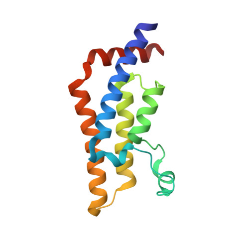
Observed bromodomain flexibility reveals histone peptide- and small molecule ligand-compatible forms of ATAD2.

Poncet-Montange, G.Zhan, Y.Bardenhagen, J.P.Petrocchi, A.Leo, E.Shi, X.Lee, G.R.Leonard, P.G.Geck Do, M.K.Cardozo, M.G.Andersen, J.N.Palmer, W.S.Jones, P.Ladbury, J.E.

(2015) Biochem J 466: 337-346

  • DOI: https://doi.org/10.1042/BJ20140933
  • Primary Citation Related Structures: 
    4TT2, 4TT4, 4TT6, 4TTE, 4TU4, 4TU6

  • PubMed Abstract: 

    Preventing histone recognition by bromodomains emerges as an attractive therapeutic approach in cancer. Overexpression of ATAD2 (ATPase family AAA domain-containing 2 isoform A) in cancer cells is associated with poor prognosis making the bromodomain of ATAD2 a promising epigenetic therapeutic target. In the development of an in vitro assay and identification of small molecule ligands, we conducted structure-guided studies which revealed a conformationally flexible ATAD2 bromodomain. Structural studies on apo-, peptide-and small molecule-ATAD2 complexes (by co-crystallization) revealed that the bromodomain adopts a 'closed', histone-compatible conformation and a more 'open' ligand-compatible conformation of the binding site respectively. An unexpected conformational change of the conserved asparagine residue plays an important role in driving the peptide-binding conformation remodelling. We also identified dimethylisoxazole-containing ligands as ATAD2 binders which aided in the validation of the in vitro screen and in the analysis of these conformational studies.


  • Organizational Affiliation
    • *Institute for Applied Cancer Science, The University of Texas MD Anderson Cancer Center, Unit 1954, 1515 Holcombe Blvd, Houston, TX 77030, U.S.A.

Macromolecule Content 

  • Total Structure Weight: 31.85 kDa 
  • Atom Count: 2,309 
  • Modeled Residue Count: 265 
  • Deposited Residue Count: 265 
  • Unique protein chains: 2

Macromolecules

Find similar proteins by:|  3D Structure
Entity ID: 1
MoleculeChains  Sequence LengthOrganismDetailsImage
ATPase family AAA domain-containing protein 2
A, B
130Homo sapiensMutation(s): 0 
Gene Names: ATAD2L16PRO2000
EC: 3.6.1.3 (PDB Primary Data), 3.6.1 (UniProt)
UniProt & NIH Common Fund Data Resources
Find proteins for Q6PL18 (Homo sapiens)
Explore Q6PL18 
Go to UniProtKB:  Q6PL18
PHAROS:  Q6PL18
GTEx:  ENSG00000156802 
Entity Groups
Sequence Clusters30% Identity50% Identity70% Identity90% Identity95% Identity100% Identity
UniProt GroupQ6PL18
Sequence Annotations
Expand
Reference Sequence
Find similar proteins by:  Sequence   |   3D Structure  
Entity ID: 2
MoleculeChains  Sequence LengthOrganismDetailsImage
Histone H3(1-21)K4AcC [auth P]5Homo sapiensMutation(s): 0 

Experimental Data & Validation

Experimental Data

  • Method: X-RAY DIFFRACTION
  • Resolution: 2.70 Å
  • R-Value Free:  0.240 (Depositor), 0.255 (DCC) 
  • R-Value Work:  0.215 (Depositor), 0.230 (DCC) 
  • R-Value Observed: 0.216 (Depositor) 
Space Group: C 2 2 21
Unit Cell:
Length ( Å )Angle ( ˚ )
a = 108.16α = 90
b = 115.68β = 90
c = 77.5γ = 90
Software Package:
Software NamePurpose
REFMACrefinement
Aimlessdata scaling
PDB_EXTRACTdata extraction

Structure Validation

View Full Validation Report


Entry History 

Revision History  (Full details and data files)

  • Version 1.0: 2014-12-24
    Type: Initial release
  • Version 1.1: 2015-03-04
    Changes: Database references
  • Version 2.0: 2022-04-13
    Changes: Advisory, Atomic model, Data collection, Database references, Derived calculations, Other, Refinement description, Source and taxonomy, Structure summary
  • Version 2.1: 2023-09-27
    Changes: Data collection, Refinement description
  • Version 2.2: 2023-11-15
    Changes: Data collection
  • Version 2.3: 2024-11-20
    Changes: Structure summary