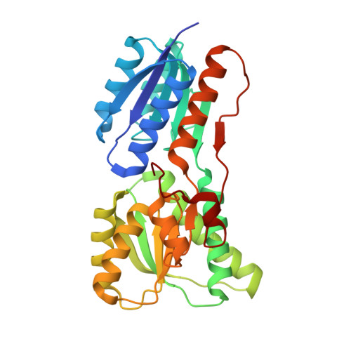
Crystal structure of carbohydrate transporter solute binding protein VEIS_2079 from Verminephrobacter eiseniae
Patskovsky, Y., Toro, R., Bhosle, R., Al Obaidi, N., Chamala, S., Scott Glenn, A., Attonito, J.D., Chowdhury, S., Lafleur, J., Siedel, R.D., Morisco, L.L., Wasserman, S.R., Hillerich, B., Love, J., Whalen, K.L., Gerlt, J.A., Almo, S.C.To be published.