4RTD

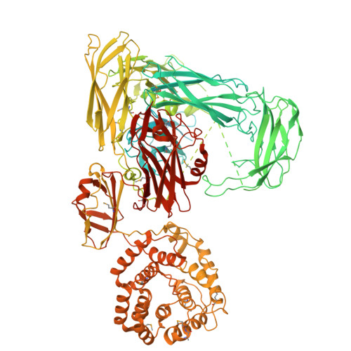
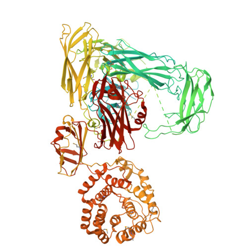
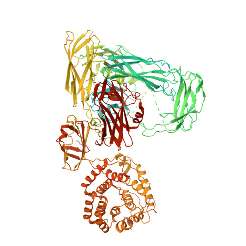
Escherichia coli alpha-2-macroglobulin activated by porcine elastase


Experimental Data Snapshot

  • Method: X-RAY DIFFRACTION
  • Resolution: 3.65 Å
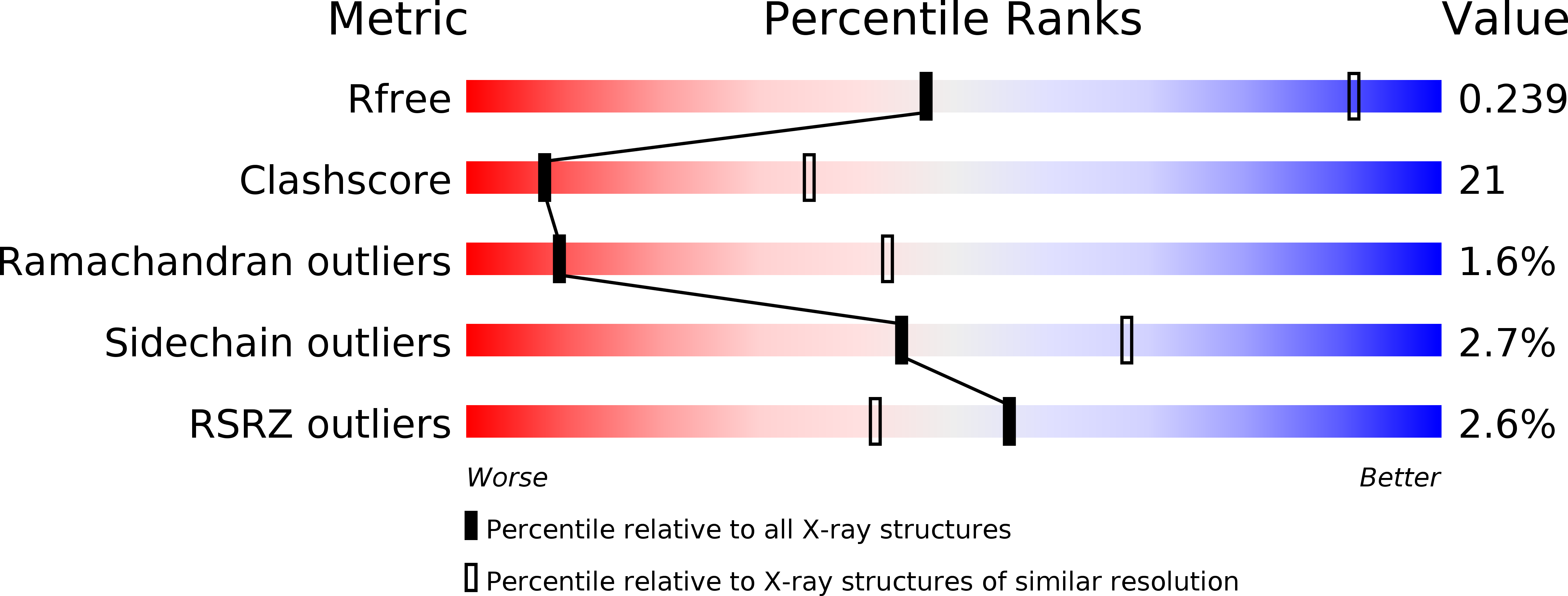
  • R-Value Free: 0.238 
  • R-Value Work: 0.177 
  • R-Value Observed: 0.180 

wwPDB Validation   3D Report Full Report


This is version 1.2 of the entry. See complete history


Literature

Structure of protease-cleaved Escherichia coli alpha-2-macroglobulin reveals a putative mechanism of conformational activation for protease entrapment.

Fyfe, C.D.Grinter, R.Josts, I.Mosbahi, K.Roszak, A.W.Cogdell, R.J.Wall, D.M.Burchmore, R.J.Byron, O.Walker, D.

(2015) Acta Crystallogr D Biol Crystallogr 71: 1478-1486

  • DOI: https://doi.org/10.1107/S1399004715008548
  • Primary Citation of Related Structures:  
    4RTD

  • PubMed Abstract: 

    Bacterial α-2-macroglobulins have been suggested to function in defence as broad-spectrum inhibitors of host proteases that breach the outer membrane. Here, the X-ray structure of protease-cleaved Escherichia coli α-2-macroglobulin is described, which reveals a putative mechanism of activation and conformational change essential for protease inhibition. In this competitive mechanism, protease cleavage of the bait-region domain results in the untethering of an intrinsically disordered region of this domain which disrupts native interdomain interactions that maintain E. coli α-2-macroglobulin in the inactivated form. The resulting global conformational change results in entrapment of the protease and activation of the thioester bond that covalently links to the attacking protease. Owing to the similarity in structure and domain architecture of Escherichia coli α-2-macroglobulin and human α-2-macroglobulin, this protease-activation mechanism is likely to operate across the diverse members of this group.


  • Organizational Affiliation

    Institute of Infection, Immunity and Inflammation, College of Medical, Veterinary and Life Sciences, University of Glasgow, Glasgow G12 8QQ, Scotland.


Macromolecules
Find similar proteins by:  (by identity cutoff)  |  3D Structure
Entity ID: 1
MoleculeChains Sequence LengthOrganismDetailsImage
Uncharacterized lipoprotein YfhM1,639Escherichia coli K-12Mutation(s): 3 
Gene Names: b2520JW2504yfhM
UniProt
Find proteins for P76578 (Escherichia coli (strain K12))
Explore P76578 
Go to UniProtKB:  P76578
Entity Groups  
Sequence Clusters30% Identity50% Identity70% Identity90% Identity95% Identity100% Identity
UniProt GroupP76578
Sequence Annotations
Expand
  • Reference Sequence
Small Molecules
Modified Residues  1 Unique
IDChains TypeFormula2D DiagramParent
MSE
Query on MSE
A
L-PEPTIDE LINKINGC5 H11 N O2 SeMET
Experimental Data & Validation

Experimental Data

  • Method: X-RAY DIFFRACTION
  • Resolution: 3.65 Å
  • R-Value Free: 0.238 
  • R-Value Work: 0.177 
  • R-Value Observed: 0.180 
  • Space Group: H 3
Unit Cell:
Length ( Å )Angle ( ˚ )
a = 176.065α = 90
b = 176.065β = 90
c = 161.131γ = 120
Software Package:
Software NamePurpose
EDNAdata collection
AutoSolphasing
REFMACrefinement
XDSdata reduction
Aimlessdata scaling

Structure Validation

View Full Validation Report


Entry History 

Deposition Data

Revision History  (Full details and data files)

  • Version 1.0: 2015-07-15
    Type: Initial release
  • Version 1.1: 2015-07-29
    Changes: Database references
  • Version 1.2: 2024-11-20
    Changes: Data collection, Database references, Derived calculations, Structure summary