4RSJ | pdb_00004rsj

Pyrococcus furiosus Smc hinge domain with an extended coiled coil


Experimental Data Snapshot

  • Method: X-RAY DIFFRACTION
  • Resolution: 3.50 Å
  • R-Value Free: 
    0.284 (Depositor), 0.293 (DCC) 
  • R-Value Work: 
    0.236 (Depositor), 0.251 (DCC) 

Starting Model: experimental
View more details

wwPDB Validation 3D Report Full Report

Validation slider image for 4RSJ

This is version 1.2 of the entry. See complete history

Literature

Molecular Basis for SMC Rod Formation and Its Dissolution upon DNA Binding.

Soh, Y.M.Burmann, F.Shin, H.C.Oda, T.Jin, K.S.Toseland, C.P.Kim, C.Lee, H.Kim, S.J.Kong, M.S.Durand-Diebold, M.L.Kim, Y.G.Kim, H.M.Lee, N.K.Sato, M.Oh, B.H.Gruber, S.

(2015) Mol Cell 57: 290-303

  • DOI: https://doi.org/10.1016/j.molcel.2014.11.023
  • Primary Citation Related Structures: 
    4RSI, 4RSJ

  • PubMed Abstract: 

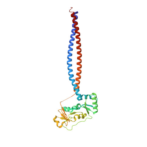
    SMC condensin complexes are central modulators of chromosome superstructure in all branches of life. Their SMC subunits form a long intramolecular coiled coil, which connects a constitutive "hinge" dimerization domain with an ATP-regulated "head" dimerization module. Here, we address the structural arrangement of the long coiled coils in SMC complexes. We unequivocally show that prokaryotic Smc-ScpAB, eukaryotic condensin, and possibly also cohesin form rod-like structures, with their coiled coils being closely juxtaposed and accurately anchored to the hinge. Upon ATP-induced binding of DNA to the hinge, however, Smc switches to a more open configuration. Our data suggest that a long-distance structural transition is transmitted from the Smc head domains to regulate Smc-ScpAB's association with DNA. These findings uncover a conserved architectural theme in SMC complexes, provide a mechanistic basis for Smc's dynamic engagement with chromosomes, and offer a molecular explanation for defects in Cornelia de Lange syndrome.


  • Organizational Affiliation
    • Department of Biological Sciences, KAIST Institute for the Biocentury, Cancer Metastasis Control Center, Korea Advanced Institute of Science and Technology, Daejeon 305-701, Korea.

Macromolecule Content 

  • Total Structure Weight: 123.44 kDa 
  • Atom Count: 7,715 
  • Modeled Residue Count: 1,064 
  • Deposited Residue Count: 1,104 
  • Unique protein chains: 1

Macromolecules

Find similar proteins by:|  3D Structure
Entity ID: 1
MoleculeChains  Sequence LengthOrganismDetailsImage
Chromosome partition protein Smc
A, B, C, D
276Pyrococcus furiosus DSM 3638Mutation(s): 0 
Gene Names: smcPF1843
UniProt
Find proteins for Q8TZY2 (Pyrococcus furiosus (strain ATCC 43587 / DSM 3638 / JCM 8422 / Vc1))
Explore Q8TZY2 
Go to UniProtKB:  Q8TZY2
Entity Groups
Sequence Clusters30% Identity50% Identity70% Identity90% Identity95% Identity100% Identity
UniProt GroupQ8TZY2
Sequence Annotations
Expand
Reference Sequence

Experimental Data & Validation

Experimental Data

  • Method: X-RAY DIFFRACTION
  • Resolution: 3.50 Å
  • R-Value Free:  0.284 (Depositor), 0.293 (DCC) 
  • R-Value Work:  0.236 (Depositor), 0.251 (DCC) 
Space Group: P 21 21 21
Unit Cell:
Length ( Å )Angle ( ˚ )
a = 101.923α = 90
b = 116.882β = 90
c = 145.493γ = 90
Software Package:
Software NamePurpose
HKL-2000data collection
PHENIXmodel building
CNSrefinement
HKL-2000data reduction
HKL-2000data scaling
PHENIXphasing

Structure Validation

View Full Validation Report


Entry History 

Deposition Data

Revision History  (Full details and data files)

  • Version 1.0: 2014-12-31
    Type: Initial release
  • Version 1.1: 2015-02-11
    Changes: Database references
  • Version 1.2: 2023-09-20
    Changes: Data collection, Database references, Refinement description