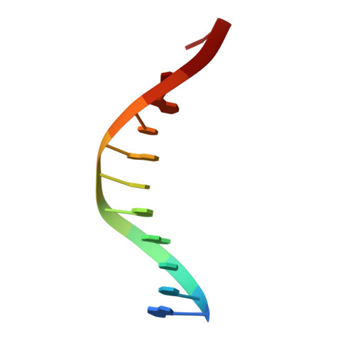
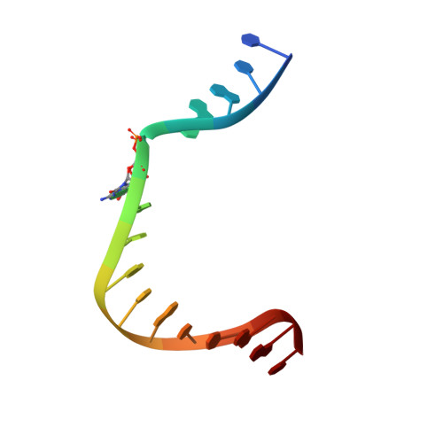
RCSB PDB - 4RQ1: Human DNA Polymerase Beta With Gapped DNA Containing an 8-oxo-7,8-dihydro-Guanine(8-oxoG) and dCTP soaked with MgCl2 for 1hr

 4RQ1

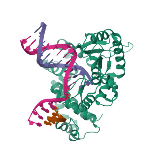
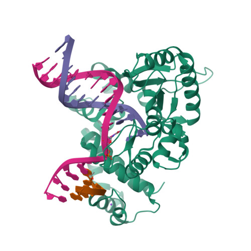
Human DNA Polymerase Beta With Gapped DNA Containing an 8-oxo-7,8-dihydro-Guanine(8-oxoG) and dCTP soaked with MgCl2 for 1hr


Experimental Data Snapshot

  • Method: X-RAY DIFFRACTION
  • Resolution: 2.70 Å
  • R-Value Free: 0.280 
  • R-Value Work: 0.203 
  • R-Value Observed: 0.207 

Starting Model: experimental
View more details

wwPDB Validation   3D Report Full Report


This is version 1.2 of the entry. See complete history


Literature

Viewing Human DNA Polymerase beta Faithfully and Unfaithfully Bypass an Oxidative Lesion by Time-Dependent Crystallography.

Vyas, R.Reed, A.J.Tokarsky, E.J.Suo, Z.

(2015) J Am Chem Soc 137: 5225-5230

  • DOI: https://doi.org/10.1021/jacs.5b02109
  • Primary Citation of Related Structures:  
    4RPX, 4RPY, 4RPZ, 4RQ0, 4RQ1, 4RQ2, 4RQ3, 4RQ4, 4RQ5, 4RQ6, 4RQ7, 4RQ8

  • PubMed Abstract: 

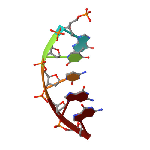
    One common oxidative DNA lesion, 8-oxo-7,8-dihydro-2'-deoxyguanine (8-oxoG), is highly mutagenic in vivo due to its anti-conformation forming a Watson-Crick base pair with correct deoxycytidine 5'-triphosphate (dCTP) and its syn-conformation forming a Hoogsteen base pair with incorrect deoxyadenosine 5'-triphosphate (dATP). Here, we utilized time-resolved X-ray crystallography to follow 8-oxoG bypass by human DNA polymerase β (hPolβ). In the 12 solved structures, both Watson-Crick (anti-8-oxoG:anti-dCTP) and Hoogsteen (syn-8-oxoG:anti-dATP) base pairing were clearly visible and were maintained throughout the chemical reaction. Additionally, a third Mg(2+) appeared during the process of phosphodiester bond formation and was located between the reacting α- and β-phosphates of the dNTP, suggesting its role in stabilizing reaction intermediates. After phosphodiester bond formation, hPolβ reopened its conformation, pyrophosphate was released, and the newly incorporated primer 3'-terminal nucleotide stacked, rather than base paired, with 8-oxoG. These structures provide the first real-time pictures, to our knowledge, of how a polymerase correctly and incorrectly bypasses a DNA lesion.


  • Organizational Affiliation

    †Department of Chemistry and Biochemistry, ‡The Ohio State Biochemistry and §Biophysics Programs, The Ohio State University, Columbus, Ohio 43210, United States.


Macromolecules

Find similar proteins by:  (by identity cutoff)  |  3D Structure
Entity ID: 1
MoleculeChains Sequence LengthOrganismDetailsImage
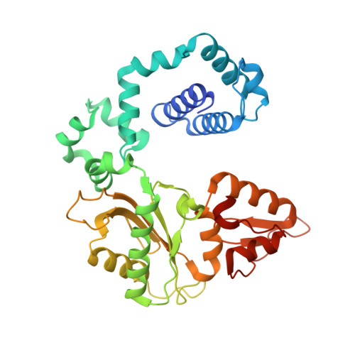
DNA polymerase beta343Homo sapiensMutation(s): 0 
Gene Names: POL BPOLB
EC: 2.7.7.7 (PDB Primary Data), 4.2.99 (PDB Primary Data), 4.2.99.18 (UniProt)
UniProt & NIH Common Fund Data Resources
Find proteins for P06746 (Homo sapiens)
Explore P06746 
Go to UniProtKB:  P06746
PHAROS:  P06746
GTEx:  ENSG00000070501 
Entity Groups  
Sequence Clusters30% Identity50% Identity70% Identity90% Identity95% Identity100% Identity
UniProt GroupP06746
Sequence Annotations
Expand
  • Reference Sequence
Experimental Data & Validation

Experimental Data

  • Method: X-RAY DIFFRACTION
  • Resolution: 2.70 Å
  • R-Value Free: 0.280 
  • R-Value Work: 0.203 
  • R-Value Observed: 0.207 
  • Space Group: P 1 21 1
Unit Cell:
Length ( Å )Angle ( ˚ )
a = 55.42α = 90
b = 80.3β = 110.27
c = 55.8γ = 90
Software Package:
Software NamePurpose
PHASERphasing
REFMACrefinement
MOSFLMdata reduction
SCALEPACKdata scaling

Structure Validation

View Full Validation Report


Entry History 

Deposition Data

Revision History  (Full details and data files)

  • Version 1.0: 2015-04-15
    Type: Initial release
  • Version 1.1: 2015-05-06
    Changes: Database references
  • Version 1.2: 2023-09-20
    Changes: Data collection, Database references, Derived calculations, Refinement description