4RMV

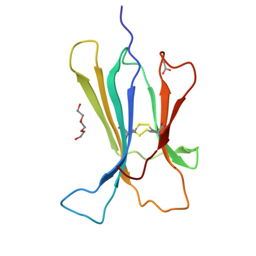
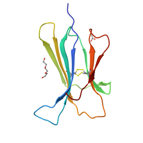
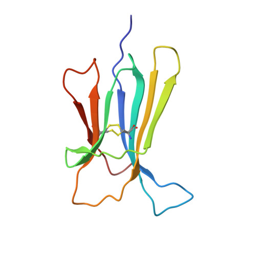
Crystal structure of the D76H Beta-2 Microglobulin mutant


Experimental Data Snapshot

  • Method: X-RAY DIFFRACTION
  • Resolution: 1.46 Å
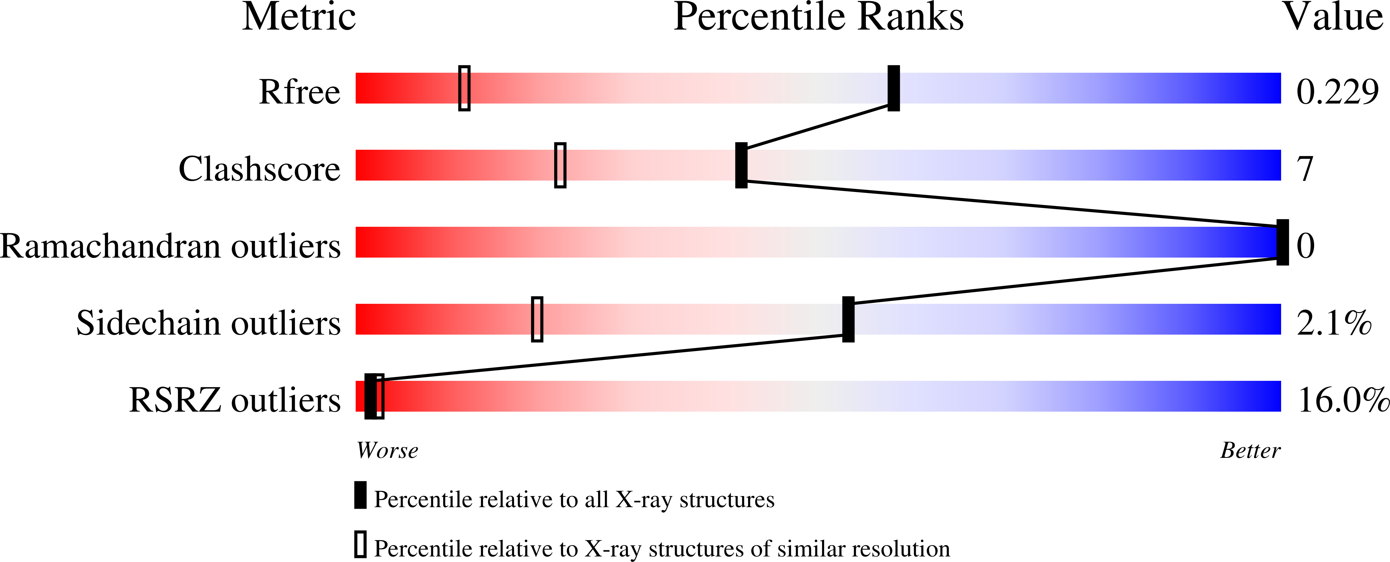
  • R-Value Free: 0.229 
  • R-Value Work: 0.186 
  • R-Value Observed: 0.190 

Starting Model: experimental
View more details

wwPDB Validation   3D Report Full Report


This is version 1.3 of the entry. See complete history


Literature

Conformational dynamics in crystals reveal the molecular bases for D76N beta-2 microglobulin aggregation propensity.

Le Marchand, T.de Rosa, M.Salvi, N.Sala, B.M.Andreas, L.B.Barbet-Massin, E.Sormanni, P.Barbiroli, A.Porcari, R.Sousa Mota, C.de Sanctis, D.Bolognesi, M.Emsley, L.Bellotti, V.Blackledge, M.Camilloni, C.Pintacuda, G.Ricagno, S.

(2018) Nat Commun 9: 1658-1658

  • DOI: https://doi.org/10.1038/s41467-018-04078-y
  • Primary Citation of Related Structures:  
    4RMU, 4RMV, 4RMW, 5CS7, 5CSB, 5CSG

  • PubMed Abstract: 

    Spontaneous aggregation of folded and soluble native proteins in vivo is still a poorly understood process. A prototypic example is the D76N mutant of beta-2 microglobulin (β2m) that displays an aggressive aggregation propensity. Here we investigate the dynamics of β2m by X-ray crystallography, solid-state NMR, and molecular dynamics simulations to unveil the effects of the D76N mutation. Taken together, our data highlight the presence of minor disordered substates in crystalline β2m. The destabilization of the outer strands of D76N β2m accounts for the increased aggregation propensity. Furthermore, the computational modeling reveals a network of interactions with residue D76 as a keystone: this model allows predicting the stability of several point mutants. Overall, our study shows how the study of intrinsic dynamics in crystallo can provide crucial answers on protein stability and aggregation propensity. The comprehensive approach here presented may well be suited for the study of other folded amyloidogenic proteins.


  • Organizational Affiliation

    Centre de RMN à Très Hauts Champs, Institut des Sciences Analytiques (UMR 5280 CNRS/UCB Lyon 1/ENS Lyon), Université de Lyon, 69100, Villeurbanne, France.


Macromolecules
Find similar proteins by:  (by identity cutoff)  |  3D Structure
Entity ID: 1
MoleculeChains Sequence LengthOrganismDetailsImage
Beta-2-microglobulin100Homo sapiensMutation(s): 1 
Gene Names: B2MCDABP0092HDCMA22PNM_004048
UniProt & NIH Common Fund Data Resources
Find proteins for P61769 (Homo sapiens)
Explore P61769 
Go to UniProtKB:  P61769
PHAROS:  P61769
GTEx:  ENSG00000166710 
Entity Groups  
Sequence Clusters30% Identity50% Identity70% Identity90% Identity95% Identity100% Identity
UniProt GroupP61769
Sequence Annotations
Expand
  • Reference Sequence
Experimental Data & Validation

Experimental Data

  • Method: X-RAY DIFFRACTION
  • Resolution: 1.46 Å
  • R-Value Free: 0.229 
  • R-Value Work: 0.186 
  • R-Value Observed: 0.190 
  • Space Group: C 1 2 1
Unit Cell:
Length ( Å )Angle ( ˚ )
a = 77.54α = 90
b = 28.871β = 125.78
c = 53.28γ = 90
Software Package:
Software NamePurpose
MxCuBEdata collection
PHASERphasing
PHENIXrefinement
MOSFLMdata reduction
SCALAdata scaling

Structure Validation

View Full Validation Report


Entry History 

Deposition Data

Revision History  (Full details and data files)

  • Version 1.0: 2015-11-18
    Type: Initial release
  • Version 1.1: 2018-06-27
    Changes: Data collection, Database references, Derived calculations
  • Version 1.2: 2023-09-20
    Changes: Data collection, Database references, Derived calculations, Refinement description
  • Version 1.3: 2024-10-30
    Changes: Structure summary