4RIB

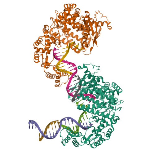
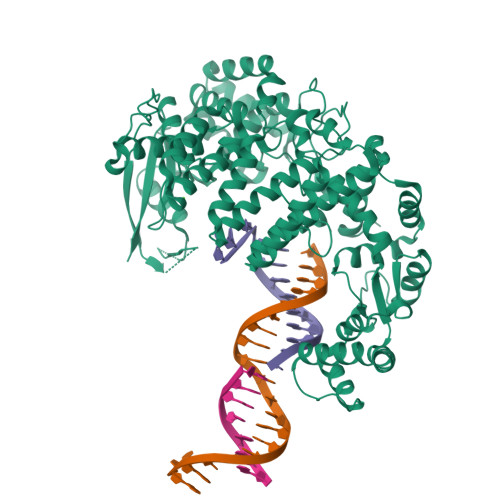
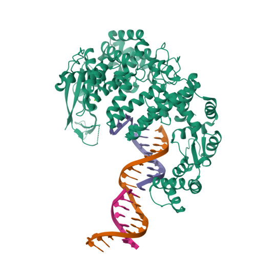
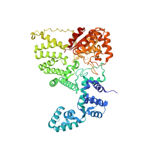
FAN1 Nuclease bound to 5' phosphorylated p(dT) single flap DNA


Experimental Data Snapshot

  • Method: X-RAY DIFFRACTION
  • Resolution: 3.25 Å
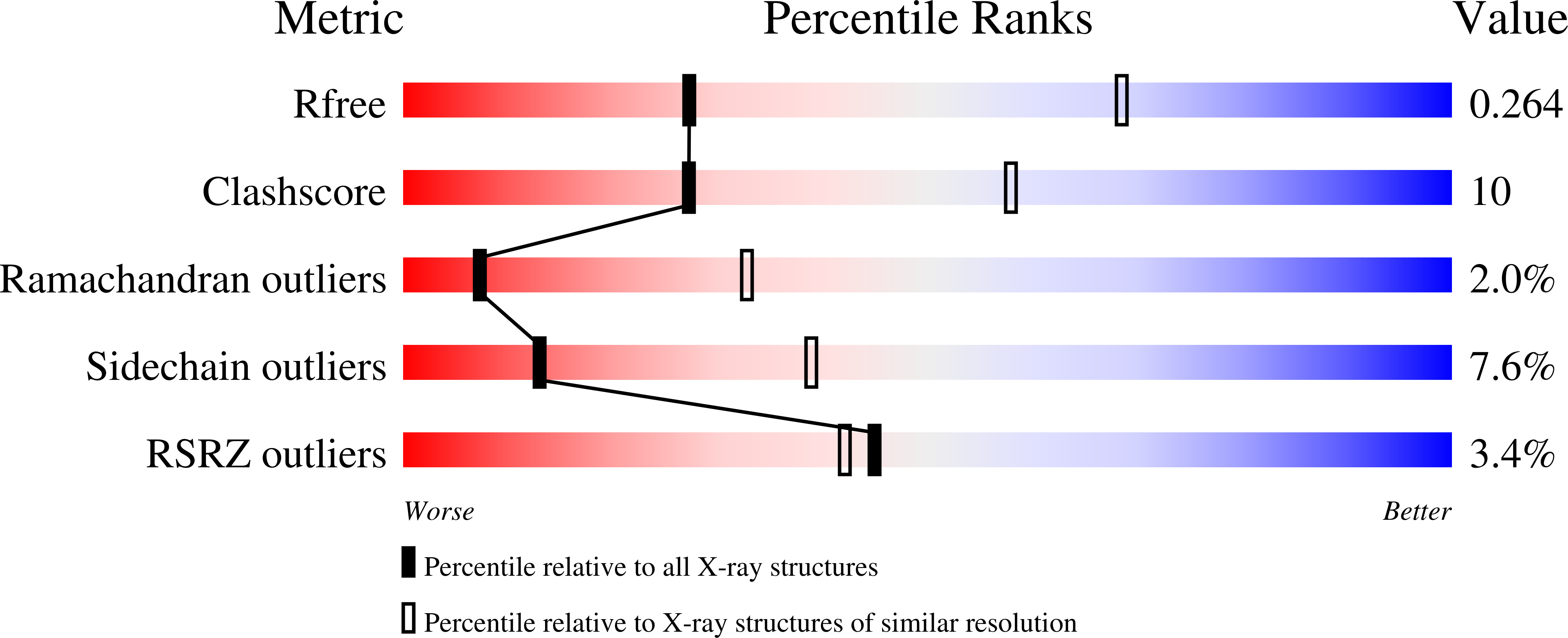
  • R-Value Free: 0.265 
  • R-Value Work: 0.226 
  • R-Value Observed: 0.227 

wwPDB Validation   3D Report Full Report


This is version 1.2 of the entry. See complete history


Literature

DNA repair. Mechanism of DNA interstrand cross-link processing by repair nuclease FAN1.

Wang, R.Persky, N.S.Yoo, B.Ouerfelli, O.Smogorzewska, A.Elledge, S.J.Pavletich, N.P.

(2014) Science 346: 1127-1130

  • DOI: https://doi.org/10.1126/science.1258973
  • Primary Citation of Related Structures:  
    4RI8, 4RI9, 4RIA, 4RIB, 4RIC, 4RID

  • PubMed Abstract: 

    DNA interstrand cross-links (ICLs) are highly toxic lesions associated with cancer and degenerative diseases. ICLs can be repaired by the Fanconi anemia (FA) pathway and through FA-independent processes involving the FAN1 nuclease. In this work, FAN1-DNA crystal structures and biochemical data reveal that human FAN1 cleaves DNA successively at every third nucleotide. In vitro, this exonuclease mechanism allows FAN1 to excise an ICL from one strand through flanking incisions. DNA access requires a 5'-terminal phosphate anchor at a nick or a 1- or 2-nucleotide flap and is augmented by a 3' flap, suggesting that FAN1 action is coupled to DNA synthesis or recombination. FAN1's mechanism of ICL excision is well suited for processing other localized DNA adducts as well.


  • Organizational Affiliation

    Structural Biology Program and Howard Hughes Medical Institute, Memorial Sloan Kettering Cancer Center, New York, NY 10065, USA.


Macromolecules

Find similar proteins by:  (by identity cutoff)  |  3D Structure
Entity ID: 1
MoleculeChains Sequence LengthOrganismDetailsImage
Fanconi-associated nuclease 1
A, B
651Homo sapiensMutation(s): 1 
Gene Names: FAN1KIAA1018MTMR15
EC: 3.1.21 (PDB Primary Data), 3.1.4.1 (PDB Primary Data)
UniProt & NIH Common Fund Data Resources
Find proteins for Q9Y2M0 (Homo sapiens)
Explore Q9Y2M0 
Go to UniProtKB:  Q9Y2M0
PHAROS:  Q9Y2M0
GTEx:  ENSG00000198690 
Entity Groups  
Sequence Clusters30% Identity50% Identity70% Identity90% Identity95% Identity100% Identity
UniProt GroupQ9Y2M0
Sequence Annotations
Expand
  • Reference Sequence

Find similar nucleic acids by:  Sequence   |   3D Structure  

Entity ID: 2
MoleculeChains LengthOrganismImage
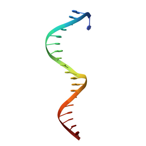
DNA (5'-D(P*TP*AP*GP*CP*CP*AP*CP*GP*CP*CP*TP*AP*GP*AP*CP*TP*CP*CP*TP*C)-3')C [auth X],
D [auth U]
20N/A
Sequence Annotations
Expand
  • Reference Sequence

Find similar nucleic acids by:  Sequence   |   3D Structure  

Entity ID: 3
MoleculeChains LengthOrganismImage
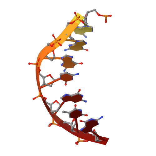
DNA (5'-D(*GP*CP*TP*GP*AP*GP*GP*AP*GP*TP*CP*T)-3')E [auth Y],
F [auth V]
12N/A
Sequence Annotations
Expand
  • Reference Sequence

Find similar nucleic acids by:  Sequence   |   3D Structure  

Entity ID: 4
MoleculeChains LengthOrganismImage
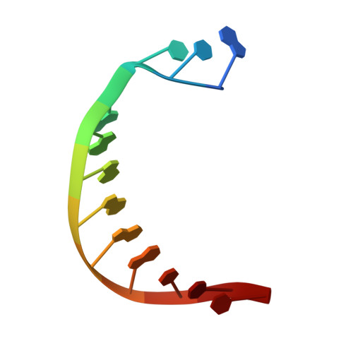
DNA (5'-D(*TP*TP*TP*TP*TP*TP*GP*AP*GP*GP*CP*GP*TP*G)-3')G [auth Z],
H [auth W]
14N/A
Sequence Annotations
Expand
  • Reference Sequence
Experimental Data & Validation

Experimental Data

  • Method: X-RAY DIFFRACTION
  • Resolution: 3.25 Å
  • R-Value Free: 0.265 
  • R-Value Work: 0.226 
  • R-Value Observed: 0.227 
  • Space Group: P 1 21 1
Unit Cell:
Length ( Å )Angle ( ˚ )
a = 97.697α = 90
b = 110.989β = 103.75
c = 105.427γ = 90
Software Package:
Software NamePurpose
SCALEPACKdata scaling
REFMACrefinement
PDB_EXTRACTdata extraction
ADSCdata collection
DENZOdata reduction
SHARPphasing

Structure Validation

View Full Validation Report


Entry History 

Deposition Data

Revision History  (Full details and data files)

  • Version 1.0: 2014-12-03
    Type: Initial release
  • Version 1.1: 2014-12-10
    Changes: Database references
  • Version 1.2: 2024-02-28
    Changes: Data collection, Database references, Derived calculations