4REP | pdb_00004rep

Crystal Structure of gamma-carotenoid desaturase


Experimental Data Snapshot

  • Method: X-RAY DIFFRACTION
  • Resolution: 1.97 Å
  • R-Value Free: 
    0.224 (Depositor), 0.226 (DCC) 
  • R-Value Work: 
    0.160 (Depositor), 0.169 (DCC) 
  • R-Value Observed: 
    0.163 (Depositor) 

wwPDB Validation 3D Report Full Report

Validation slider image for 4REP

Ligand Structure Quality Assessment 


This is version 1.2 of the entry. See complete history

Literature

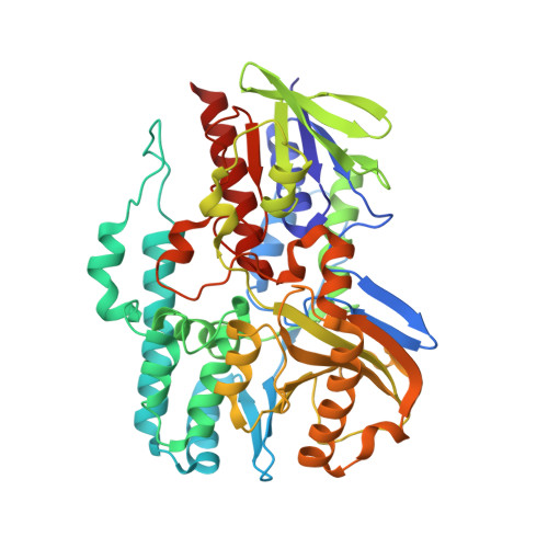
Crystal structure of 1'-OH-carotenoid 3,4-desaturase from Nonlabens dokdonensis DSW-6.

Ahn, J.W.Kim, K.J.

(2015) Enzyme Microb Technol 77: 29-37

  • DOI: https://doi.org/10.1016/j.enzmictec.2015.05.005
  • Primary Citation Related Structures: 
    4REP

  • PubMed Abstract: 

    The γ-carotenoids, such as myxol and saproxanthin, have a high potential to be utilized in nutraceutical and pharmaceutical industries for their neuro-protective and antioxidant effects. CrtD is involved in the production of γ-carotenoids by desaturating the C3'-C4' position of 1'-OH-γ-carotenoid. We determined the crystal structure of CrtD from Nonlabens dokdonensis DSW-6 (NdCrtD), the first structure of CrtD family enzymes. The NdCrtD structure was composed of two distinct domains, an FAD-binding domain and a substrate-binding domain, and the substrate-binding domain can be divided into two subdomains, a Rossmann fold-like subdomain and a lid subdomain. Although the FAD-binding domain showed a structure similar to canonical FAD-containing enzymes, the substrate-binding domain exhibited a novel structure to constitute a long and hydrophobic tunnel with a length of ∼40 Å. The molecular docking-simulation reveals that the tunnel provides an appropriate substrate-binding site for the carotenoid such as 1'-OH-γ-carotene with a length of ∼35 Å. We could predict residues related to recognize the 1'-hydroxyl group and to stabilize the hydrophobic end without hydroxyl group. Moreover, we suggest that the flexible entrance loop may undergo an open-closed formational change during the binding of the substrate.


  • Organizational Affiliation
    • School of Life Sciences, KNU Creative BioResearch Group, Kyungpook National University, Daehak-ro 80, Buk-ku, Daegu 702-701, South Korea.

Macromolecule Content 

  • Total Structure Weight: 56.79 kDa 
  • Atom Count: 4,221 
  • Modeled Residue Count: 489 
  • Deposited Residue Count: 495 
  • Unique protein chains: 1

Macromolecules

Find similar proteins by:|  3D Structure
Entity ID: 1
MoleculeChains  Sequence LengthOrganismDetailsImage
Gamma-carotene desaturase495Nonlabens dokdonensis DSW-6Mutation(s): 0 
Gene Names: DDD_2394
EC: 1.3.99 (PDB Primary Data), 1.3.99.27 (UniProt)
UniProt
Find proteins for L7WC64 (Nonlabens dokdonensis (strain DSM 17205 / KCTC 12402 / DSW-6))
Explore L7WC64 
Go to UniProtKB:  L7WC64
Entity Groups
Sequence Clusters30% Identity50% Identity70% Identity90% Identity95% Identity100% Identity
UniProt GroupL7WC64
Sequence Annotations
Expand
Reference Sequence

Small Molecules

Experimental Data & Validation

Experimental Data

  • Method: X-RAY DIFFRACTION
  • Resolution: 1.97 Å
  • R-Value Free:  0.224 (Depositor), 0.226 (DCC) 
  • R-Value Work:  0.160 (Depositor), 0.169 (DCC) 
  • R-Value Observed: 0.163 (Depositor) 
Space Group: P 1 21 1
Unit Cell:
Length ( Å )Angle ( ˚ )
a = 48.369α = 90
b = 72.835β = 110.29
c = 68.137γ = 90
Software Package:
Software NamePurpose
DENZOdata reduction
SCALEPACKdata scaling
REFMACrefinement
PDB_EXTRACTdata extraction
HKL-2000data collection
HKL-2000data reduction
SOLVEphasing

Structure Validation

View Full Validation Report


Ligand Structure Quality Assessment 


Entry History 

Deposition Data

Revision History  (Full details and data files)

  • Version 1.0: 2015-07-15
    Type: Initial release
  • Version 1.1: 2017-11-22
    Changes: Refinement description
  • Version 1.2: 2024-02-28
    Changes: Data collection, Database references, Derived calculations