4R8A | pdb_00004r8a

Crystal structure of paFAN1 - 5' flap DNA complex


Experimental Data Snapshot

  • Method: X-RAY DIFFRACTION
  • Resolution: 3.20 Å
  • R-Value Free: 
    0.234 (Depositor), 0.241 (DCC) 
  • R-Value Work: 
    0.196 (Depositor), 0.205 (DCC) 
  • R-Value Observed: 
    0.197 (Depositor) 

wwPDB Validation 3D Report Full Report

Validation slider image for 4R8A

This is version 1.1 of the entry. See complete history

Literature

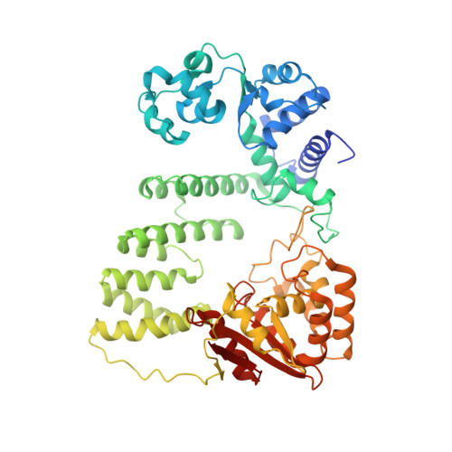
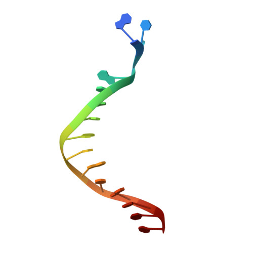
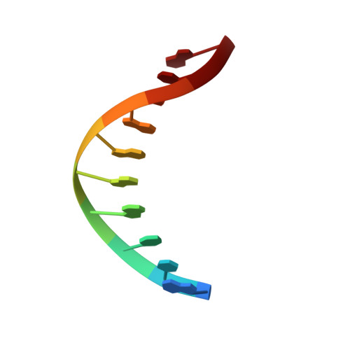
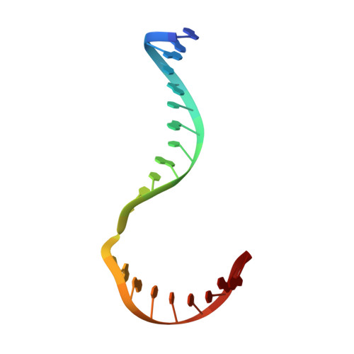
Crystal structure of a Fanconi anemia-associated nuclease homolog bound to 5' flap DNA: basis of interstrand cross-link repair by FAN1

Gwon, G.H.Kim, Y.R.Liu, Y.Watson, A.T.Jo, A.Etheridge, T.J.Yuan, F.Zhang, Y.Kim, Y.C.Carr, A.M.Cho, Y.

(2014) Genes Dev 28: 2276-2290

  • DOI: https://doi.org/10.1101/gad.248492.114
  • Primary Citation Related Structures: 
    4R89, 4R8A

  • PubMed Abstract: 

    Fanconi anemia (FA) is an autosomal recessive genetic disorder caused by defects in any of 15 FA genes responsible for processing DNA interstrand cross-links (ICLs). The ultimate outcome of the FA pathway is resolution of cross-links, which requires structure-selective nucleases. FA-associated nuclease 1 (FAN1) is believed to be recruited to lesions by a monoubiquitinated FANCI-FANCD2 (ID) complex and participates in ICL repair. Here, we determined the crystal structure of Pseudomonas aeruginosa FAN1 (PaFAN1) lacking the UBZ (ubiquitin-binding zinc) domain in complex with 5' flap DNA. All four domains of the right-hand-shaped PaFAN1 are involved in DNA recognition, with each domain playing a specific role in bending DNA at the nick. The six-helix bundle that binds the junction connects to the catalytic viral replication and repair (VRR) nuclease (VRR nuc) domain, enabling FAN1 to incise the scissile phosphate a few bases distant from the junction. The six-helix bundle also inhibits the cleavage of intact Holliday junctions. PaFAN1 shares several conserved features with other flap structure-selective nucleases despite structural differences. A clamping motion of the domains around the wedge helix, which acts as a pivot, facilitates nucleolytic cleavage. The PaFAN1 structure provides insights into how archaeal Holliday junction resolvases evolved to incise 5' flap substrates and how FAN1 integrates with the FA complex to participate in ICL repair.


  • Organizational Affiliation
    • Department of Life Science, Pohang University of Science and Technology, Pohang 790-784, South Korea;

Macromolecule Content 

  • Total Structure Weight: 157.34 kDa 
  • Atom Count: 10,652 
  • Modeled Residue Count: 1,168 
  • Deposited Residue Count: 1,210 
  • Unique protein chains: 1
  • Unique nucleic acid chains: 3

Macromolecules


Find similar proteins by:|  3D Structure
Entity ID: 1
MoleculeChains  Sequence LengthOrganismDetailsImage
Uncharacterized proteinA,
E [auth F]
559Pseudomonas aeruginosa PAO1Mutation(s): 0 
Gene Names: PA1865
EC: 3.1.4.1
UniProt
Find proteins for Q9I2N0 (Pseudomonas aeruginosa (strain ATCC 15692 / DSM 22644 / CIP 104116 / JCM 14847 / LMG 12228 / 1C / PRS 101 / PAO1))
Explore Q9I2N0 
Go to UniProtKB:  Q9I2N0
Entity Groups
Sequence Clusters30% Identity50% Identity70% Identity90% Identity95% Identity100% Identity
UniProt GroupQ9I2N0
Sequence Annotations
Expand
Reference Sequence
Find similar nucleic acids by:  Sequence
Entity ID: 2
MoleculeChains LengthOrganismImage
DNA (5'-D(P*AP*CP*CP*AP*GP*AP*CP*AP*CP*AP*CP*AP*TP*TP*C)-3')B,
F [auth G]
15N/A
Sequence Annotations
Expand
Reference Sequence
Find similar nucleic acids by:  Sequence
Entity ID: 3
MoleculeChains LengthOrganismImage
DNA (5'-D(P*GP*TP*TP*GP*GP*GP*AP*TP*TP*G)-3')C,
G [auth H]
10N/A
Sequence Annotations
Expand
Reference Sequence
Find similar nucleic acids by:  Sequence
Entity ID: 4
MoleculeChains LengthOrganismImage
DNA (5'-D(P*GP*AP*AP*TP*GP*TP*GP*TP*GP*TP*CP*TP*CP*AP*AP*TP*CP*CP*CP*AP*A)-3')D,
H [auth I]
21N/A
Sequence Annotations
Expand
Reference Sequence

Experimental Data & Validation

Experimental Data

  • Method: X-RAY DIFFRACTION
  • Resolution: 3.20 Å
  • R-Value Free:  0.234 (Depositor), 0.241 (DCC) 
  • R-Value Work:  0.196 (Depositor), 0.205 (DCC) 
  • R-Value Observed: 0.197 (Depositor) 
Space Group: P 1 21 1
Unit Cell:
Length ( Å )Angle ( ˚ )
a = 76.581α = 90
b = 106.663β = 89.97
c = 142.18γ = 90
Software Package:
Software NamePurpose
HKL-2000data collection
PHASERphasing
PHENIXrefinement
HKL-2000data reduction
HKL-2000data scaling

Structure Validation

View Full Validation Report


Entry History 

Deposition Data

Revision History  (Full details and data files)

  • Version 1.0: 2014-10-29
    Type: Initial release
  • Version 1.1: 2024-03-20
    Changes: Data collection, Database references