


Crystal structure of a Quinonprotein alcohol dehydrogenase-like protein (BT1487) from Bacteroides thetaiotaomicron VPI-5482 at 2.64 A resolution
Joint Center for Structural Genomics (JCSG)To be published.
Experimental Data Snapshot
wwPDB Validation 3D Report Full Report

Entity ID: 1 | |||||
|---|---|---|---|---|---|
| Molecule | Chains | Sequence Length | Organism | Details | Image |
| Quinonprotein alcohol dehydrogenase-like protein | 427 | Bacteroides thetaiotaomicron VPI-5482 | Mutation(s): 0 Gene Names: BT_1487 | 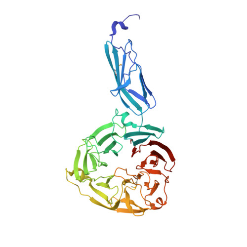 | |
UniProt | |||||
Entity Groups | |||||
| Sequence Clusters | 30% Identity50% Identity70% Identity90% Identity95% Identity100% Identity | ||||
| UniProt Group | Q8A7N7 | ||||
Sequence AnnotationsExpand | |||||
Reference Sequence | |||||
| Ligands 4 Unique | |||||
|---|---|---|---|---|---|
| ID | Chains | Name / Formula / InChI Key | 2D Diagram | 3D Interactions | |
| 7PE Download:Ideal Coordinates CCD File | AA [auth D] E [auth A] F [auth A] G [auth A] O [auth B] | 2-(2-(2-(2-(2-(2-ETHOXYETHOXY)ETHOXY)ETHOXY)ETHOXY)ETHOXY)ETHANOL C14 H30 O7 UKXKPKBTMYNOFS-UHFFFAOYSA-N |  | ||
| ACT Download:Ideal Coordinates CCD File | H [auth A], I [auth A], T [auth B], U [auth B] | ACETATE ION C2 H3 O2 QTBSBXVTEAMEQO-UHFFFAOYSA-M |  | ||
| CA Download:Ideal Coordinates CCD File | J [auth A], K [auth A], L [auth A], M [auth A], V [auth B] | CALCIUM ION Ca BHPQYMZQTOCNFJ-UHFFFAOYSA-N |  | ||
| CL Download:Ideal Coordinates CCD File | N [auth A], Z [auth C] | CHLORIDE ION Cl VEXZGXHMUGYJMC-UHFFFAOYSA-M |  | ||
| Modified Residues 1 Unique | |||||
|---|---|---|---|---|---|
| ID | Chains | Type | Formula | 2D Diagram | Parent |
| MSE Query on MSE | A, B, C, D | L-PEPTIDE LINKING | C5 H11 N O2 Se |  | MET |
| Length ( Å ) | Angle ( ˚ ) |
|---|---|
| a = 155.545 | α = 90 |
| b = 155.545 | β = 90 |
| c = 94.635 | γ = 120 |
| Software Name | Purpose |
|---|---|
| MolProbity | model building |
| PDB_EXTRACT | data extraction |
| SHELX | phasing |
| SHARP | phasing |
| XSCALE | data scaling |
| BUSTER-TNT | refinement |
| XDS | data reduction |
| SHELXD | phasing |
| BUSTER | refinement |


