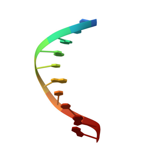
4R4A

Racemic crystal structure of a cobalt-bound B-DNA duplex


Experimental Data Snapshot

  • Method: X-RAY DIFFRACTION
  • Resolution: 1.49 Å
  • R-Value Free: 0.233 
  • R-Value Work: 0.209 
  • R-Value Observed: 0.210 

Starting Model: experimental
View more details

wwPDB Validation   3D Report Full Report


Ligand Structure Quality Assessment 


This is version 1.2 of the entry. See complete history


Literature

Racemic DNA crystallography.

Mandal, P.K.Collie, G.W.Kauffmann, B.Huc, I.

(2014) Angew Chem Int Ed Engl 53: 14424-14427

  • DOI: https://doi.org/10.1002/anie.201409014
  • Primary Citation of Related Structures:  
    4R44, 4R45, 4R47, 4R48, 4R49, 4R4A, 4R4D

  • PubMed Abstract: 

    Racemates increase the chances of crystallization by allowing molecular contacts to be formed in a greater number of ways. With the advent of protein synthesis, the production of protein racemates and racemic-protein crystallography are now possible. Curiously, racemic DNA crystallography had not been investigated despite the commercial availability of L- and D-deoxyribo-oligonucleotides. Here, we report a study into racemic DNA crystallography showing the strong propensity of racemic DNA mixtures to form racemic crystals. We describe racemic crystal structures of various DNA sequences and folded conformations, including duplexes, quadruplexes, and a four-way junction, showing that the advantages of racemic crystallography should extend to DNA.


  • Organizational Affiliation

    Université de Bordeaux, CBMN, UMR5248, Institut Européen de Chimie et Biologie, 2 rue Robert Escarpit, 33600 Pessac (France); CNRS, CBMN, UMR5248.


Macromolecules

Find similar nucleic acids by:  Sequence   |   3D Structure  

Entity ID: 1
MoleculeChains LengthOrganismImage
5'-D(*CP*CP*GP*GP*TP*AP*CP*CP*GP*G)-3'
A, B
10synthetic construct
Sequence Annotations
Expand
  • Reference Sequence
Experimental Data & Validation

Experimental Data

  • Method: X-RAY DIFFRACTION
  • Resolution: 1.49 Å
  • R-Value Free: 0.233 
  • R-Value Work: 0.209 
  • R-Value Observed: 0.210 
  • Space Group: P -1
Unit Cell:
Length ( Å )Angle ( ˚ )
a = 26.286α = 62.25
b = 32.352β = 73.14
c = 33.5γ = 68.56
Software Package:
Software NamePurpose
DNAdata collection
PHASERphasing
REFMACrefinement
XDSdata reduction
d*TREKdata scaling

Structure Validation

View Full Validation Report


Ligand Structure Quality Assessment 


Entry History 

Deposition Data

Revision History  (Full details and data files)

  • Version 1.0: 2014-11-12
    Type: Initial release
  • Version 1.1: 2015-01-14
    Changes: Database references
  • Version 1.2: 2023-09-20
    Changes: Data collection, Database references, Derived calculations, Refinement description