4R3E | pdb_00004r3e

Structure of the spliceosomal peptidyl-prolyl cis-trans isomerase Cwc27 from Homo sapiens


Experimental Data Snapshot

  • Method: X-RAY DIFFRACTION
  • Resolution: 2.00 Å
  • R-Value Free: 
    0.217 (Depositor), 0.184 (DCC) 
  • R-Value Work: 
    0.172 (Depositor) 
  • R-Value Observed: 
    0.174 (Depositor) 

Starting Model: experimental
View more details

wwPDB Validation 3D Report Full Report

Validation slider image for 4R3E

This is version 1.3 of the entry. See complete history

Literature

Structure and evolution of the spliceosomal peptidyl-prolyl cis-trans isomerase Cwc27.

Ulrich, A.Wahl, M.C.

(2014) Acta Crystallogr D Biol Crystallogr 70: 3110-3123

  • DOI: https://doi.org/10.1107/S1399004714021695
  • Primary Citation Related Structures: 
    4R3E, 4R3F

  • PubMed Abstract: 

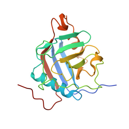
    Cwc27 is a spliceosomal cyclophilin-type peptidyl-prolyl cis-trans isomerase (PPIase). Here, the crystal structure of a relatively protease-resistant N-terminal fragment of human Cwc27 containing the PPIase domain was determined at 2.0 Å resolution. The fragment exhibits a C-terminal appendix and resides in a reduced state compared with the previous oxidized structure of a similar fragment. By combining multiple sequence alignments spanning the eukaryotic tree of life and secondary-structure prediction, Cwc27 proteins across the entire eukaryotic kingdom were identified. This analysis revealed the specific loss of a crucial active-site residue in higher eukaryotic Cwc27 proteins, suggesting that the protein evolved from a prolyl isomerase to a pure proline binder. Noting a fungus-specific insertion in the PPIase domain, the 1.3 Å resolution crystal structure of the PPIase domain of Cwc27 from Chaetomium thermophilum was also determined. Although structurally highly similar in the core domain, the C. thermophilum protein displayed a higher thermal stability than its human counterpart, presumably owing to the combined effect of several amino-acid exchanges that reduce the number of long side chains with strained conformations and create new intramolecular interactions, in particular increased hydrogen-bond networks.


  • Organizational Affiliation
    • Laboratory of Structural Biochemistry, Freie Universität Berlin, Takustrasse 6, 14195 Berlin, Germany.

Macromolecule Content 

  • Total Structure Weight: 20.41 kDa 
  • Atom Count: 1,588 
  • Modeled Residue Count: 173 
  • Deposited Residue Count: 180 
  • Unique protein chains: 1

Macromolecules

Find similar proteins by:|  3D Structure
Entity ID: 1
MoleculeChains  Sequence LengthOrganismDetailsImage
Peptidyl-prolyl cis-trans isomerase CWC27 homolog180Homo sapiensMutation(s): 0 
Gene Names: CWC27SDCCAG10UNQ438/PRO871
EC: 5.2.1.8
UniProt & NIH Common Fund Data Resources
Find proteins for Q6UX04 (Homo sapiens)
Explore Q6UX04 
Go to UniProtKB:  Q6UX04
PHAROS:  Q6UX04
GTEx:  ENSG00000153015 
Entity Groups
Sequence Clusters30% Identity50% Identity70% Identity90% Identity95% Identity100% Identity
UniProt GroupQ6UX04
Sequence Annotations
Expand
Reference Sequence

Experimental Data & Validation

Experimental Data

  • Method: X-RAY DIFFRACTION
  • Resolution: 2.00 Å
  • R-Value Free:  0.217 (Depositor), 0.184 (DCC) 
  • R-Value Work:  0.172 (Depositor) 
  • R-Value Observed: 0.174 (Depositor) 
Space Group: P 65
Unit Cell:
Length ( Å )Angle ( ˚ )
a = 85.86α = 90
b = 85.86β = 90
c = 55.02γ = 120
Software Package:
Software NamePurpose
MAR345dtbdata collection
PHASERphasing
REFMACrefinement
XDSdata reduction
XSCALEdata scaling

Structure Validation

View Full Validation Report


Entry History 

Deposition Data

Revision History  (Full details and data files)

  • Version 1.0: 2014-11-19
    Type: Initial release
  • Version 1.1: 2014-12-10
    Changes: Database references
  • Version 1.2: 2014-12-24
    Changes: Database references
  • Version 1.3: 2023-09-20
    Changes: Data collection, Database references, Derived calculations, Refinement description