4R1A | pdb_00004r1a

Crystal Structure of FVO strain Plasmodium falciparum AMA1


Experimental Data Snapshot

  • Method: X-RAY DIFFRACTION
  • Resolution: 2.00 Å
  • R-Value Free: 
    0.263 (Depositor), 0.260 (DCC) 
  • R-Value Work: 
    0.199 (Depositor), 0.204 (DCC) 
  • R-Value Observed: 
    0.203 (Depositor) 

Starting Model: experimental
View more details

wwPDB Validation 3D Report Full Report

Validation slider image for 4R1A

This is version 1.2 of the entry. See complete history

Literature

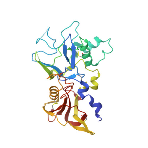
Structure and Dynamics of Apical Membrane Antigen 1 from Plasmodium falciparum FVO.

Lim, S.S.Yang, W.Krishnarjuna, B.Kannan Sivaraman, K.Chandrashekaran, I.R.Kass, I.MacRaild, C.A.Devine, S.M.Debono, C.O.Anders, R.F.Scanlon, M.J.Scammells, P.J.Norton, R.S.McGowan, S.

(2014) Biochemistry 53: 7310-7320

  • DOI: https://doi.org/10.1021/bi5012089
  • Primary Citation Related Structures: 
    4R19, 4R1A, 4R1B, 4R1C

  • PubMed Abstract: 

    Apical membrane antigen 1 (AMA1) interacts with RON2 to form a protein complex that plays a key role in the invasion of host cells by malaria parasites. Blocking this protein-protein interaction represents a potential route to controlling malaria and related parasitic diseases, but the polymorphic nature of AMA1 has proven to be a major challenge to vaccine-induced antibodies and peptide inhibitors exerting strain-transcending inhibitory effects. Here we present the X-ray crystal structure of AMA1 domains I and II from Plasmodium falciparum strain FVO. We compare our new structure to those of AMA1 from P. falciparum 3D7 and Plasmodium vivax. A combination of normalized B factor analysis and computational methods has been used to investigate the flexibility of the domain I loops and how this correlates with their roles in determining the strain specificity of human antibody responses and inhibitory peptides. We also investigated the domain II loop, a key region involved in inhibitor binding, by comparison of multiple AMA1 crystal structures. Collectively, these results provide valuable insights that should contribute to the design of strain-transcending agents targeting P. falciparum AMA1.


  • Organizational Affiliation
    • Medicinal Chemistry, Monash Institute of Pharmaceutical Sciences, Monash University , Parkville, Victoria 3052, Australia.

Macromolecule Content 

  • Total Structure Weight: 38.33 kDa 
  • Atom Count: 2,174 
  • Modeled Residue Count: 265 
  • Deposited Residue Count: 335 
  • Unique protein chains: 1

Macromolecules

Find similar proteins by:|  3D Structure
Entity ID: 1
MoleculeChains  Sequence LengthOrganismDetailsImage
Apical membrane antigen 1335Plasmodium falciparum FCR-3/GambiaMutation(s): 0 
Gene Names: AMA-1PF83
UniProt
Find proteins for P50490 (Plasmodium falciparum (isolate FCR-3 / Gambia))
Explore P50490 
Go to UniProtKB:  P50490
Entity Groups
Sequence Clusters30% Identity50% Identity70% Identity90% Identity95% Identity100% Identity
UniProt GroupP50490
Sequence Annotations
Expand
Reference Sequence

Experimental Data & Validation

Experimental Data

  • Method: X-RAY DIFFRACTION
  • Resolution: 2.00 Å
  • R-Value Free:  0.263 (Depositor), 0.260 (DCC) 
  • R-Value Work:  0.199 (Depositor), 0.204 (DCC) 
  • R-Value Observed: 0.203 (Depositor) 
Space Group: C 1 2 1
Unit Cell:
Length ( Å )Angle ( ˚ )
a = 109.794α = 90
b = 37.788β = 94.04
c = 71.706γ = 90
Software Package:
Software NamePurpose
ADSCdata collection
PHASESphasing
PHENIXrefinement
MOSFLMdata reduction
SCALAdata scaling

Structure Validation

View Full Validation Report


Entry History 

Deposition Data

Revision History  (Full details and data files)

  • Version 1.0: 2015-01-14
    Type: Initial release
  • Version 1.1: 2023-09-20
    Changes: Data collection, Database references, Refinement description
  • Version 1.2: 2024-11-20
    Changes: Structure summary