4R09 | pdb_00004r09

Crystal structure of human TLR8 in complex with ORN06S


Experimental Data Snapshot

  • Method: X-RAY DIFFRACTION
  • Resolution: 2.62 Å
  • R-Value Free: 
    0.285 (Depositor), 0.282 (DCC) 
  • R-Value Work: 
    0.216 (Depositor), 0.217 (DCC) 
  • R-Value Observed: 
    0.220 (Depositor) 

wwPDB Validation 3D Report Full Report

Validation slider image for 4R09

Ligand Structure Quality Assessment 


This is version 2.1 of the entry. See complete history

Literature

Toll-like receptor 8 senses degradation products of single-stranded RNA.

Tanji, H.Ohto, U.Shibata, T.Taoka, M.Yamauchi, Y.Isobe, T.Miyake, K.Shimizu, T.

(2015) Nat Struct Mol Biol 22: 109-115

  • DOI: https://doi.org/10.1038/nsmb.2943
  • Primary Citation Related Structures: 
    4R07, 4R08, 4R09, 4R0A

  • PubMed Abstract: 

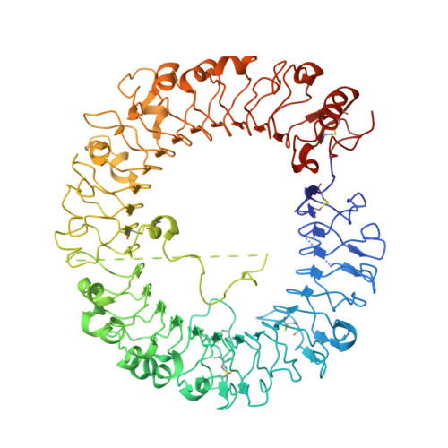
    Toll-like receptor 8 (TLR8) recognizes viral or bacterial single-stranded RNA (ssRNA) and activates innate immune systems. TLR8 is activated by uridine- and guanosine-rich ssRNA as well as by certain synthetic chemicals; however, the molecular basis for ssRNA recognition has remained unknown. In this study, to elucidate the recognition mechanism of ssRNA, we determined the crystal structures of human TLR8 in complex with ssRNA. TLR8 recognized two degradation products of ssRNA—uridine and a short oligonucleotide—at two distinct sites: uridine bound the site on the dimerization interface where small chemical ligands are recognized, whereas short oligonucleotides bound a newly identified site on the concave surface of the TLR8 horseshoe structure. Site-directed mutagenesis revealed that both binding sites were essential for activation of TLR8 by ssRNA. These results demonstrate that TLR8 is a sensor for both uridine and a short oligonucleotide derived from RNA.


  • Organizational Affiliation
    • Graduate School of Pharmaceutical Sciences, University of Tokyo, Tokyo, Japan.

Macromolecule Content 

  • Total Structure Weight: 383.59 kDa 
  • Atom Count: 24,895 
  • Modeled Residue Count: 2,993 
  • Deposited Residue Count: 3,244 
  • Unique protein chains: 1

Macromolecules

Find similar proteins by:|  3D Structure
Entity ID: 1
MoleculeChains  Sequence LengthOrganismDetailsImage
Toll-like receptor 8
A, B, C, D
811Homo sapiensMutation(s): 0 
Gene Names: TLR8UNQ249/PRO286
UniProt & NIH Common Fund Data Resources
Find proteins for Q9NR97 (Homo sapiens)
Explore Q9NR97 
Go to UniProtKB:  Q9NR97
PHAROS:  Q9NR97
GTEx:  ENSG00000101916 
Entity Groups
Sequence Clusters30% Identity50% Identity70% Identity90% Identity95% Identity100% Identity
UniProt GroupQ9NR97
Glycosylation
Glycosylation Sites: 3Go to GlyGen: Q9NR97-1
Sequence Annotations
Expand
Reference Sequence

Oligosaccharides

Help  
Entity ID: 2
MoleculeChains Length2D Diagram GlycosylationD Interactions
alpha-D-mannopyranose-(1-3)-[alpha-D-mannopyranose-(1-6)]beta-D-mannopyranose-(1-4)-2-acetamido-2-deoxy-beta-D-glucopyranose-(1-4)-2-acetamido-2-deoxy-beta-D-glucopyranose
E, K
5N-Glycosylation
Glycosylation Resources
GlyTouCan: G22768VO
GlyCosmos: G22768VO
GlyGen: G22768VO
Entity ID: 3
MoleculeChains Length2D Diagram GlycosylationD Interactions
2-acetamido-2-deoxy-beta-D-glucopyranose-(1-4)-2-acetamido-2-deoxy-beta-D-glucopyranose
F, G, I, J, M
F, G, I, J, M, O
2N-Glycosylation
Glycosylation Resources
GlyTouCan: G42666HT
GlyCosmos: G42666HT
GlyGen: G42666HT
Entity ID: 4
MoleculeChains Length2D Diagram GlycosylationD Interactions
beta-D-mannopyranose-(1-4)-2-acetamido-2-deoxy-beta-D-glucopyranose-(1-4)-2-acetamido-2-deoxy-beta-D-glucopyranose
H, L, N
3N-Glycosylation
Glycosylation Resources
GlyTouCan: G15407YE
GlyCosmos: G15407YE
GlyGen: G15407YE

Small Molecules

Ligands 4 Unique
IDChains Name / Formula / InChI Key2D Diagram3D Interactions
06S

Query on 06S


Download:Ideal Coordinates CCD File
BA [auth D],
Q [auth A],
U [auth B],
X [auth C]
O-[(2R,3S,4R,5R)-5-(2-amino-6-oxo-3,6-dihydro-9H-purin-9-yl)-2-({[(S)-({(2R,3S,4R,5R)-5-(2,4-dioxo-3,4-dihydropyrimidin-1(2H)-yl)-4-hydroxy-2-[(thiophosphonooxy)methyl]tetrahydrofuran-3-yl}oxy)(sulfanyl)phosphoryl]oxy}methyl)-4-hydroxytetrahydrofuran-3-yl] dihydrogen (S)-phosphorothioate
C19 H26 N7 O16 P3 S3
PRHQISJBVGJFKN-VXGQQCRQSA-N
UPT

Query on UPT


Download:Ideal Coordinates CCD File
CA [auth D],
R [auth A],
V [auth B],
Y [auth C]
1-[(2R,3aR,4R,6R,6aR)-2-hydroxy-6-(hydroxymethyl)-2-sulfidotetrahydrofuro[3,4-d][1,3,2]dioxaphosphol-4-yl]pyrimidine-2,4(1H,3H)-dione
C9 H11 N2 O7 P S
OIDQHQCLWHMGIL-VQAIERIMSA-N
URI

Query on URI


Download:Ideal Coordinates CCD File
AA [auth D],
P [auth A],
T [auth B],
Z [auth D]
URIDINE
C9 H12 N2 O6
DRTQHJPVMGBUCF-XVFCMESISA-N
NAG

Query on NAG


Download:Ideal Coordinates CCD File
DA [auth D],
S [auth A],
W [auth B]
2-acetamido-2-deoxy-beta-D-glucopyranose
C8 H15 N O6
OVRNDRQMDRJTHS-FMDGEEDCSA-N

Experimental Data & Validation

Experimental Data

  • Method: X-RAY DIFFRACTION
  • Resolution: 2.62 Å
  • R-Value Free:  0.285 (Depositor), 0.282 (DCC) 
  • R-Value Work:  0.216 (Depositor), 0.217 (DCC) 
  • R-Value Observed: 0.220 (Depositor) 
Space Group: P 1 21 1
Unit Cell:
Length ( Å )Angle ( ˚ )
a = 86.683α = 90
b = 141.342β = 89.46
c = 170.042γ = 90
Software Package:
Software NamePurpose
MOLREPphasing
REFMACrefinement
HKL-2000data reduction
HKL-2000data scaling

Structure Validation

View Full Validation Report


Ligand Structure Quality Assessment 


Entry History 

Deposition Data

Revision History  (Full details and data files)

  • Version 1.0: 2015-01-14
    Type: Initial release
  • Version 1.1: 2015-02-18
    Changes: Database references
  • Version 2.0: 2020-07-29
    Type: Remediation
    Reason: Carbohydrate remediation
    Changes: Advisory, Atomic model, Data collection, Database references, Derived calculations, Structure summary
  • Version 2.1: 2024-11-20
    Changes: Data collection, Database references, Structure summary