4QQN | pdb_00004qqn

Protein arginine methyltransferase 3 in complex with compound MTV044246


Experimental Data Snapshot

  • Method: X-RAY DIFFRACTION
  • Resolution: 2.08 Å
  • R-Value Free: 
    0.212 (Depositor), 0.224 (DCC) 
  • R-Value Work: 
    0.183 (Depositor), 0.194 (DCC) 
  • R-Value Observed: 
    0.184 (Depositor) 

Starting Model: experimental
View more details

wwPDB Validation 3D Report Full Report

Validation slider image for 4QQN

Ligand Structure Quality Assessment 


This is version 1.2 of the entry. See complete history

Literature

Discovery of Potent and Selective Allosteric Inhibitors of Protein Arginine Methyltransferase 3 (PRMT3).

Kaniskan, H.U.Eram, M.S.Zhao, K.Szewczyk, M.M.Yang, X.Schmidt, K.Luo, X.Xiao, S.Dai, M.He, F.Zang, I.Lin, Y.Li, F.Dobrovetsky, E.Smil, D.Min, S.J.Lin-Jones, J.Schapira, M.Atadja, P.Li, E.Barsyte-Lovejoy, D.Arrowsmith, C.H.Brown, P.J.Liu, F.Yu, Z.Vedadi, M.Jin, J.

(2018) J Med Chem 61: 1204-1217

  • DOI: https://doi.org/10.1021/acs.jmedchem.7b01674
  • Primary Citation Related Structures: 
    4QQN

  • PubMed Abstract: 

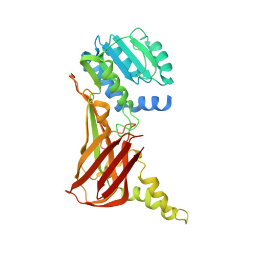
    PRMT3 catalyzes the asymmetric dimethylation of arginine residues of various proteins. It is crucial for maturation of ribosomes and has been implicated in several diseases. We recently disclosed a highly potent, selective, and cell-active allosteric inhibitor of PRMT3, compound 4. Here, we report comprehensive structure-activity relationship studies that target the allosteric binding site of PRMT3. We conducted design, synthesis, and evaluation of novel compounds in biochemical, selectivity, and cellular assays that culminated in the discovery of 4 and other highly potent (IC 50 values: ∼10-36 nM), selective, and cell-active allosteric inhibitors of PRMT3 (compounds 29, 30, 36, and 37). In addition, we generated compounds that are very close analogs of these potent inhibitors but displayed drastically reduced potency as negative controls (compounds 49-51). These inhibitors and negative controls are valuable chemical tools for the biomedical community to further investigate biological functions and disease associations of PRMT3.


  • Organizational Affiliation
    • Center for Chemical Biology and Drug Discovery, Departments of Pharmacological Sciences and Oncological Sciences, Tisch Cancer Institute, Icahn School of Medicine at Mount Sinai , New York, New York 10029, United States.

Macromolecule Content 

  • Total Structure Weight: 38.74 kDa 
  • Atom Count: 2,577 
  • Modeled Residue Count: 299 
  • Deposited Residue Count: 340 
  • Unique protein chains: 1

Macromolecules

Find similar proteins by:|  3D Structure
Entity ID: 1
MoleculeChains  Sequence LengthOrganismDetailsImage
PRMT3 protein340Homo sapiensMutation(s): 0 
Gene Names: PRMT3
EC: 2.1.1.319
UniProt & NIH Common Fund Data Resources
Find proteins for O60678 (Homo sapiens)
Explore O60678 
Go to UniProtKB:  O60678
PHAROS:  O60678
GTEx:  ENSG00000185238 
Entity Groups
Sequence Clusters30% Identity50% Identity70% Identity90% Identity95% Identity100% Identity
UniProt GroupO60678
Sequence Annotations
Expand
Reference Sequence

Small Molecules

Ligands 4 Unique
IDChains Name / Formula / InChI Key2D Diagram3D Interactions
3BQ

Query on 3BQ


Download:Ideal Coordinates CCD File
B [auth A]1-{2-[1-(aminomethyl)cyclohexyl]ethyl}-3-isoquinolin-6-ylurea
C19 H26 N4 O
NYAKHMNXWJGORW-UHFFFAOYSA-N
GOL

Query on GOL


Download:Ideal Coordinates CCD File
C [auth A]GLYCEROL
C3 H8 O3
PEDCQBHIVMGVHV-UHFFFAOYSA-N
CL

Query on CL


Download:Ideal Coordinates CCD File
D [auth A]CHLORIDE ION
Cl
VEXZGXHMUGYJMC-UHFFFAOYSA-M
UNX

Query on UNX


Download:Ideal Coordinates CCD File
E [auth A]
F [auth A]
G [auth A]
H [auth A]
I [auth A]
E [auth A],
F [auth A],
G [auth A],
H [auth A],
I [auth A],
J [auth A],
K [auth A],
L [auth A],
M [auth A],
N [auth A],
O [auth A],
P [auth A]
UNKNOWN ATOM OR ION
X
Binding Affinity Annotations 
IDSourceBinding Affinity
3BQ BindingDB:  4QQN IC50: 291 (nM) from 1 assay(s)

Experimental Data & Validation

Experimental Data

  • Method: X-RAY DIFFRACTION
  • Resolution: 2.08 Å
  • R-Value Free:  0.212 (Depositor), 0.224 (DCC) 
  • R-Value Work:  0.183 (Depositor), 0.194 (DCC) 
  • R-Value Observed: 0.184 (Depositor) 
Space Group: P 43 21 2
Unit Cell:
Length ( Å )Angle ( ˚ )
a = 70.319α = 90
b = 70.319β = 90
c = 172.918γ = 90
Software Package:
Software NamePurpose
SCALEPACKdata scaling
REFMACrefinement
PDB_EXTRACTdata extraction
HKL-3000data reduction
HKL-3000data scaling
MOLREPphasing
Cootmodel building

Structure Validation

View Full Validation Report


Ligand Structure Quality Assessment 


Entry History 

Revision History  (Full details and data files)

  • Version 1.0: 2014-09-17
    Type: Initial release
  • Version 1.1: 2018-05-16
    Changes: Data collection, Database references
  • Version 1.2: 2023-09-20
    Changes: Data collection, Database references, Derived calculations, Refinement description