4QLN | pdb_00004qln

structure of ydao riboswitch binding with c-di-dAMP


Experimental Data Snapshot

  • Method: X-RAY DIFFRACTION
  • Resolution: 2.65 Å
  • R-Value Free: 
    0.290 (Depositor), 0.290 (DCC) 
  • R-Value Work: 
    0.217 (Depositor), 0.220 (DCC) 
  • R-Value Observed: 
    0.221 (Depositor) 

wwPDB Validation   3D Report Full Report


Ligand Structure Quality Assessment 

Created with Raphaël 2.3.0Worse 01 BetterLigand structure goodness of fit to experimental dataBest fitted 2BAClick on this verticalbar to view details

This is version 1.2 of the entry. See complete history


Literature

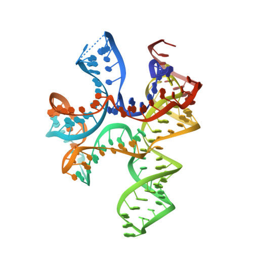
c-di-AMP binds the ydaO riboswitch in two pseudo-symmetry-related pockets.

Ren, A.Patel, D.J.

(2014) Nat Chem Biol 10: 780-786

  • DOI: https://doi.org/10.1038/nchembio.1606
  • Primary Citation of Related Structures:  
    4QLM, 4QLN

  • PubMed Abstract: 

    The ydaO riboswitch, involved in sporulation, osmotic stress responses and cell wall metabolism, targets the second messenger cyclic-di-AMP with subnanomolar affinity. We have solved the structure of c-di-AMP bound to the Thermoanaerobacter tengcongensis ydaO riboswitch, thereby identifying a five-helical scaffold containing a zippered-up bubble, a pseudoknot and long-range tertiary base pairs. Highlights include the identification of two c-di-AMP binding pockets on the same face of the riboswitch, related by pseudo-two-fold symmetry, with potential for cross-talk between sites mediated by adjacently positioned base-stacking alignments connecting pockets. The adenine rings of bound c-di-AMP molecules are wedged between bases and stabilized by stacking, base-sugar and sugar-sugar intermolecular hydrogen bonding interactions. The structural studies are complemented by isothermal titration calorimetry-based binding studies of mutants mediating key tertiary intermolecular contacts. The T. tengcongensis ydaO riboswitch, like its Bacillus subtilis counterpart, most likely functions through a transcription termination mechanism, with the c-di-AMP bound state representing an 'off' switch.


  • Organizational Affiliation

    Structural Biology Program, Memorial Sloan-Kettering Cancer Center, New York, New York, USA.


Macromolecules
Find similar nucleic acids by:  (by identity cutoff)  |  3D Structure
Entity ID: 1
MoleculeChains LengthOrganismImage
RNA (117-MER)125Caldanaerobacter subterraneus subsp. tengcongensis MB4
Sequence Annotations
Expand
  • Reference Sequence
Experimental Data & Validation

Experimental Data

  • Method: X-RAY DIFFRACTION
  • Resolution: 2.65 Å
  • R-Value Free:  0.290 (Depositor), 0.290 (DCC) 
  • R-Value Work:  0.217 (Depositor), 0.220 (DCC) 
  • R-Value Observed: 0.221 (Depositor) 
Space Group: C 2 2 21
Unit Cell:
Length ( Å )Angle ( ˚ )
a = 77.874α = 90
b = 87.051β = 90
c = 144.553γ = 90
Software Package:
Software NamePurpose
XDSdata scaling
PHASERphasing
PHENIXrefinement
XDSdata reduction

Structure Validation

View Full Validation Report


Ligand Structure Quality Assessment 

Created with Raphaël 2.3.0Worse 01 BetterLigand structure goodness of fit to experimental dataBest fitted 2BAClick on this verticalbar to view details

Entry History 

Deposition Data

Revision History  (Full details and data files)

  • Version 1.0: 2014-08-13
    Type: Initial release
  • Version 1.1: 2014-09-03
    Changes: Database references
  • Version 1.2: 2024-02-28
    Changes: Data collection, Database references, Derived calculations, Structure summary