4QCJ | pdb_00004qcj

Crystal Structure of OdhI from Corynebacterium glutamicum


Experimental Data Snapshot

  • Method: X-RAY DIFFRACTION
  • Resolution: 2.00 Å
  • R-Value Free: 
    0.240 (Depositor), 0.248 (DCC) 
  • R-Value Work: 
    0.173 (Depositor), 0.181 (DCC) 
  • R-Value Observed: 
    0.180 (Depositor) 

wwPDB Validation 3D Report Full Report

Validation slider image for 4QCJ

This is version 1.2 of the entry. See complete history

Literature

Interaction of 2-oxoglutarate dehydrogenase OdhA with its inhibitor OdhI in Corynebacterium glutamicum: Mutants and a model.

Raasch, K.Bocola, M.Labahn, J.Leitner, A.Eggeling, L.Bott, M.

(2014) J Biotechnol 191: 99-105

  • DOI: https://doi.org/10.1016/j.jbiotec.2014.05.023
  • Primary Citation Related Structures: 
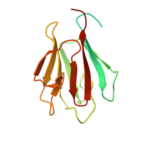
    4QCJ

  • PubMed Abstract: 

    Pyruvate dehydrogenase and oxoglutarate dehydrogenase catalyze key reactions in central metabolism. In Corynebacterium glutamicum and related bacteria like Mycobacterium tuberculosis both activities reside in a novel protein supercomplex with the fusion protein OdhA catalyzing the conversion of oxoglutarate to succinyl-coenzyme A. This activity is inhibited by the forkhead-associated (FHA) domain of the small autoinhibitory protein OdhI. Here we used a biological screen which enabled us to isolate suppressor mutants that are influenced in OdhA-OdhI interaction. Five mutants carrying an OdhI mutation were isolated and one with an OdhA mutation. The OdhA mutein OdhA-C704E and three additional C704 variants were constructed. They exhibited unaltered or even slightly enhanced OdhA activity but showed reduced inhibition and interaction with OdhI. The FHA domain of OdhI was crystallized and its structure found in full agreement with previously determined NMR structures. Based on further structural studies, OdhA-OdhI crosslinking experiments, and modeling we discuss the experimental data generated on OdhA-OdhI interaction, with the latter protein representing a rare example of an FHA domain also recognizing a non-phosphorylated interaction partner.


  • Organizational Affiliation
    • Institute of Bio- and Geosciences, IBG-1: Biotechnology, Forschungszentrum Jülich, Jülich, Germany.

Macromolecule Content 

  • Total Structure Weight: 15.42 kDa 
  • Atom Count: 912 
  • Modeled Residue Count: 101 
  • Deposited Residue Count: 143 
  • Unique protein chains: 1

Macromolecules

Find similar proteins by:|  3D Structure
Entity ID: 1
MoleculeChains  Sequence LengthOrganismDetailsImage
Oxoglutarate dehydrogenase inhibitor143Corynebacterium glutamicum ATCC 13032Mutation(s): 0 
Gene Names: cg1630Cgl1441odhI
UniProt
Find proteins for Q8NQJ3 (Corynebacterium glutamicum (strain ATCC 13032 / DSM 20300 / JCM 1318 / BCRC 11384 / CCUG 27702 / LMG 3730 / NBRC 12168 / NCIMB 10025 / NRRL B-2784 / 534))
Explore Q8NQJ3 
Go to UniProtKB:  Q8NQJ3
Entity Groups
Sequence Clusters30% Identity50% Identity70% Identity90% Identity95% Identity100% Identity
UniProt GroupQ8NQJ3
Sequence Annotations
Expand
Reference Sequence

Experimental Data & Validation

Experimental Data

  • Method: X-RAY DIFFRACTION
  • Resolution: 2.00 Å
  • R-Value Free:  0.240 (Depositor), 0.248 (DCC) 
  • R-Value Work:  0.173 (Depositor), 0.181 (DCC) 
  • R-Value Observed: 0.180 (Depositor) 
Space Group: P 21 21 2
Unit Cell:
Length ( Å )Angle ( ˚ )
a = 51.026α = 90
b = 51.229β = 90
c = 35.52γ = 90
Software Package:
Software NamePurpose
ADSCdata collection
MOLREPphasing
PHENIXrefinement
MOSFLMdata reduction
SCALAdata scaling

Structure Validation

View Full Validation Report


Entry History 

Deposition Data

Revision History  (Full details and data files)

  • Version 1.0: 2014-07-02
    Type: Initial release
  • Version 1.1: 2014-12-31
    Changes: Database references
  • Version 1.2: 2024-02-28
    Changes: Data collection, Database references