4QBN | pdb_00004qbn

VRR_NUC domain


Experimental Data Snapshot

  • Method: X-RAY DIFFRACTION
  • Resolution: 1.85 Å
  • R-Value Free: 
    0.197 (Depositor), 0.199 (DCC) 
  • R-Value Work: 
    0.163 (Depositor), 0.165 (DCC) 
  • R-Value Observed: 
    0.165 (Depositor) 

wwPDB Validation 3D Report Full Report

Validation slider image for 4QBN

This is version 1.2 of the entry. See complete history

Literature

FAN1 activity on asymmetric repair intermediates is mediated by an atypical monomeric virus-type replication-repair nuclease domain.

Pennell, S.Declais, A.C.Li, J.Haire, L.F.Berg, W.Saldanha, J.W.Taylor, I.A.Rouse, J.Lilley, D.M.Smerdon, S.J.

(2014) Cell Rep 8: 84-93

  • DOI: https://doi.org/10.1016/j.celrep.2014.06.001
  • Primary Citation Related Structures: 
    4QBL, 4QBN, 4QBO

  • PubMed Abstract: 

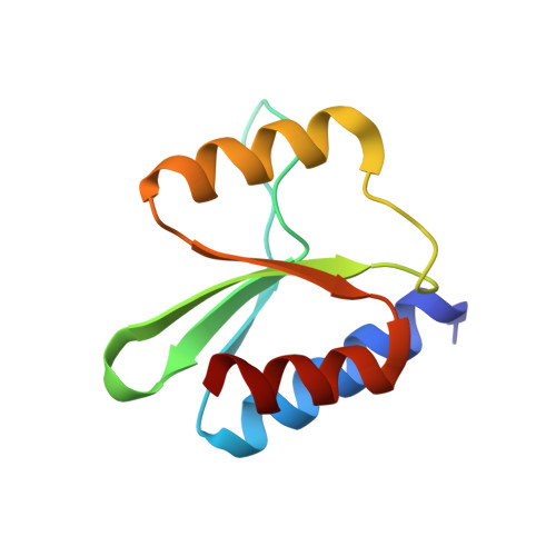
    FAN1 is a structure-selective DNA repair nuclease with 5' flap endonuclease activity, involved in the repair of interstrand DNA crosslinks. It is the only eukaryotic protein with a virus-type replication-repair nuclease ("VRR-Nuc") "module" that commonly occurs as a standalone domain in many bacteria and viruses. Crystal structures of three representatives show that they structurally resemble Holliday junction resolvases (HJRs), are dimeric in solution, and are able to cleave symmetric four-way junctions. In contrast, FAN1 orthologs are monomeric and cleave 5' flap structures in vitro, but not Holliday junctions. Modeling of the VRR-Nuc domain of FAN1 reveals that it has an insertion, which packs against the dimerization interface observed in the structures of the viral/bacterial VRR-Nuc proteins. We propose that these additional structural elements in FAN1 prevent dimerization and bias specificity toward flap structures.


  • Organizational Affiliation
    • Division of Molecular Structure, MRC National Institute for Medical Research, The Ridgeway, London NW7 1AA, UK. Electronic address: spennel@nimr.mrc.ac.uk.

Macromolecule Content 

  • Total Structure Weight: 22.27 kDa 
  • Atom Count: 1,662 
  • Modeled Residue Count: 186 
  • Deposited Residue Count: 186 
  • Unique protein chains: 1

Macromolecules

Find similar proteins by:|  3D Structure
Entity ID: 1
MoleculeChains  Sequence LengthOrganismDetailsImage
Nuclease
A, B
93Salmonella phage SETP3Mutation(s): 0 
UniProt
Find proteins for A3EZR7 (Salmonella phage SETP3)
Explore A3EZR7 
Go to UniProtKB:  A3EZR7
Entity Groups
Sequence Clusters30% Identity50% Identity70% Identity90% Identity95% Identity100% Identity
UniProt GroupA3EZR7
Sequence Annotations
Expand
Reference Sequence

Experimental Data & Validation

Experimental Data

  • Method: X-RAY DIFFRACTION
  • Resolution: 1.85 Å
  • R-Value Free:  0.197 (Depositor), 0.199 (DCC) 
  • R-Value Work:  0.163 (Depositor), 0.165 (DCC) 
  • R-Value Observed: 0.165 (Depositor) 
Space Group: F 2 2 2
Unit Cell:
Length ( Å )Angle ( ˚ )
a = 86.221α = 90
b = 99.329β = 90
c = 109.868γ = 90
Software Package:
Software NamePurpose
PHASERphasing
PHENIXrefinement
DENZOdata reduction
SCALEPACKdata scaling

Structure Validation

View Full Validation Report


Entry History 

Deposition Data

Revision History  (Full details and data files)

  • Version 1.0: 2014-09-10
    Type: Initial release
  • Version 1.1: 2017-11-22
    Changes: Refinement description
  • Version 1.2: 2024-02-28
    Changes: Data collection, Database references, Derived calculations