4QBB | pdb_00004qbb

Structure of the foot-and-mouth disease virus leader proteinase in complex with inhibitor (N~2~-[(3S)-4-({(2R)-1-[(4-CARBAMIMIDAMIDOBUTYL)AMINO]-4-METHYL-1-OXOPENTAN-2-YL}AMINO)-3-HYDROXY-4-OXOBUTANOYL]-L-ARGINYL-L-PROLINAMIDE)


Experimental Data Snapshot

  • Method: X-RAY DIFFRACTION
  • Resolution: 1.60 Å
  • R-Value Free: 
    0.201 (Depositor), 0.201 (DCC) 
  • R-Value Work: 
    0.170 (Depositor), 0.172 (DCC) 
  • R-Value Observed: 
    0.171 (Depositor) 

wwPDB Validation 3D Report Full Report

Validation slider image for 4QBB

Ligand Structure Quality Assessment 


This is version 1.1 of the entry. See complete history

Literature

Foot-and-mouth disease virus leader proteinase: Structural insights into the mechanism of intermolecular cleavage.

Steinberger, J.Grishkovskaya, I.Cencic, R.Juliano, L.Juliano, M.A.Skern, T.

(2014) Virology 468-470C: 397-408

  • DOI: https://doi.org/10.1016/j.virol.2014.08.023
  • Primary Citation Related Structures: 
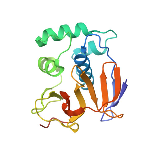
    4QBB

  • PubMed Abstract: 

    Translation of foot-and-mouth disease virus RNA initiates at one of two start codons leading to the synthesis of two forms of leader proteinase L(pro) (Lab(pro) and Lb(pro)). These forms free themselves from the viral polyprotein by intra- and intermolecular self-processing and subsequently cleave the cellular eukaryotic initiation factor (eIF) 4 G. During infection, Lb(pro) removes six residues from its own C-terminus, generating sLb(pro). We present the structure of sLb(pro) bound to the inhibitor E64-R-P-NH2, illustrating how sLb(pro) can cleave between Lys/Gly and Gly/Arg pairs. In intermolecular cleavage on polyprotein substrates, Lb(pro) was unaffected by P1 or P1' substitutions and processed a substrate containing nine eIF4GI cleavage site residues whereas sLb(pro) failed to cleave the eIF4GI containing substrate and cleaved appreciably more slowly on mutated substrates. Introduction of 70 eIF4GI residues bearing the Lb(pro) binding site restored cleavage. These data imply that Lb(pro) and sLb(pro) may have different functions in infected cells.


  • Organizational Affiliation
    • Max F. Perutz Laboratories, Medical University of Vienna, Dr. Bohr-Gasse 9/3, A-1030 Vienna, Austria.

Macromolecule Content 

  • Total Structure Weight: 59.14 kDa 
  • Atom Count: 4,440 
  • Modeled Residue Count: 472 
  • Deposited Residue Count: 501 
  • Unique protein chains: 1

Macromolecules

Find similar proteins by:|  3D Structure
Entity ID: 1
MoleculeChains  Sequence LengthOrganismDetailsImage
Leader protease
A, B, C
167Foot-and-mouth disease virus (strain O1)Mutation(s): 0 
EC: 3.4.22.46
UniProt
Find proteins for P03305 (Foot-and-mouth disease virus serotype O)
Explore P03305 
Go to UniProtKB:  P03305
Entity Groups
Sequence Clusters30% Identity50% Identity70% Identity90% Identity95% Identity100% Identity
UniProt GroupP03305
Sequence Annotations
Expand
Reference Sequence

Experimental Data & Validation

Experimental Data

  • Method: X-RAY DIFFRACTION
  • Resolution: 1.60 Å
  • R-Value Free:  0.201 (Depositor), 0.201 (DCC) 
  • R-Value Work:  0.170 (Depositor), 0.172 (DCC) 
  • R-Value Observed: 0.171 (Depositor) 
Space Group: P 1 21 1
Unit Cell:
Length ( Å )Angle ( ˚ )
a = 45.808α = 90
b = 110.682β = 98.12
c = 56.774γ = 90
Software Package:
Software NamePurpose
SCALAdata scaling
PHENIXrefinement
PDB_EXTRACTdata extraction
MxCuBEdata collection
XDSdata reduction
REFMACphasing

Structure Validation

View Full Validation Report


Ligand Structure Quality Assessment 


Entry History 

Deposition Data

Revision History  (Full details and data files)

  • Version 1.0: 2014-11-05
    Type: Initial release
  • Version 1.1: 2024-11-06
    Changes: Data collection, Database references, Derived calculations, Structure summary