Crystal structure and functional implications of LprF from Mycobacterium tuberculosis and M. bovis
Kim, J.S., Jiao, L., Oh, J.I., Ha, N.C., Kim, Y.H.(2014) Acta Crystallogr D Biol Crystallogr 70: 2619-2630
- PubMed: 25286846 Search on PubMed
- DOI: https://doi.org/10.1107/S1399004714016599
- Primary Citation Related Structures:
4QA8 - PubMed Abstract:
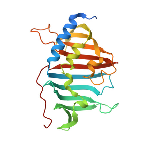
The Gram-positive bacteria Mycobacterium tuberculosis and M. bovis are causative agents of tuberculosis in humans and cattle. The lipoprotein LprF is found in M. tuberculosis and M. bovis but not in the nonpathogenic M. smegmatis. To date, the role of LprF remains to be elucidated. In this study, the crystal structure of LprF has been determined at 1.1 Å resolution. The overall structure is similar to that of a homologue, LprG, with a central hydrophobic cavity that binds a triacylated glycolipid. LprF exhibited a central cavity structure similar to that of LprG, but with a smaller cavity that binds two alkyl chains. Consistently, subsequent mass-spectrometric analysis revealed that the bound ligand was a diacylated glycolipid, as found in the structure. Furthermore, an increased ratio of lipoarabinomannan to lipomannan in the mycobacterial cell wall was observed when lprF was introduced into M. smegmatis. These observations suggested that LprF transfers the diacylated glycolipid from the plasma membrane to the cell wall, which might be related to the pathogenesis of the bacteria.
- College of Pharmacy and Research Institute for Drug Development, Pusan National University, Busan, Republic of Korea.
Organizational Affiliation: