4Q8D | pdb_00004q8d

Crystal structure of a macrocyclic beta-sheet peptide containing two beta-strands from amyloid beta residues 15-23


Experimental Data Snapshot

  • Method: X-RAY DIFFRACTION
  • Resolution: 1.75 Å
  • R-Value Free: 
    0.220 (Depositor), 0.227 (DCC) 
  • R-Value Work: 
    0.179 (Depositor), 0.182 (DCC) 
  • R-Value Observed: 
    0.186 (Depositor) 

wwPDB Validation 3D Report Full Report

Validation slider image for 4Q8D

This is version 2.0 of the entry. See complete history

Literature

A Fibril-Like Assembly of Oligomers of a Peptide Derived from beta-Amyloid.

Pham, J.D.Spencer, R.K.Chen, K.H.Nowick, J.S.

(2014) J Am Chem Soc 136: 12682-12690

  • DOI: https://doi.org/10.1021/ja505713y
  • Primary Citation Related Structures: 
    4Q8D

  • PubMed Abstract: 

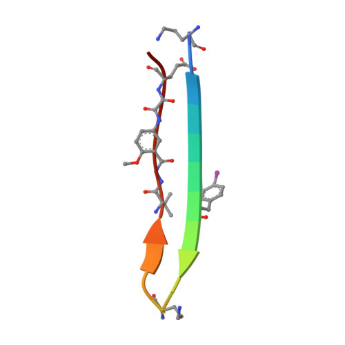
    A macrocyclic β-sheet peptide containing two nonapeptide segments based on Aβ(15-23) (QKLVFFAED) forms fibril-like assemblies of oligomers in the solid state. The X-ray crystallographic structure of macrocyclic β-sheet peptide 3 was determined at 1.75 Å resolution. The macrocycle forms hydrogen-bonded dimers, which further assemble along the fibril axis in a fashion resembling a herringbone pattern. The extended β-sheet comprising the dimers is laminated against a second layer of dimers through hydrophobic interactions to form a fibril-like assembly that runs the length of the crystal lattice. The second layer is offset by one monomer subunit, so that the fibril-like assembly is composed of partially overlapping dimers, rather than discrete tetramers. In aqueous solution, macrocyclic β-sheet 3 and homologues 4 and 5 form discrete tetramers, rather than extended fibril-like assemblies. The fibril-like assemblies of oligomers formed in the solid state by macrocyclic β-sheet 3 represent a new mode of supramolecular assembly not previously observed for the amyloidogenic central region of Aβ. The structures observed at atomic resolution for this peptide model system may offer insights into the structures of oligomers and oligomer assemblies formed by full-length Aβ and may provide a window into the propagation and replication of amyloid oligomers.


  • Organizational Affiliation
    • Department of Chemistry, University of California, Irvine , Irvine, California 92697-2025, United States.

Macromolecule Content 

  • Total Structure Weight: 5.04 kDa 
  • Atom Count: 381 
  • Modeled Residue Count: 36 
  • Deposited Residue Count: 36 
  • Unique protein chains: 1

Macromolecules

Find similar proteins by:  Sequence   |   3D Structure  
Entity ID: 1
MoleculeChains  Sequence LengthOrganismDetailsImage
macrocyclic beta-sheet peptide incorporating residues amyloid beta 15-23
A, B
18N/AMutation(s): 0 

Small Molecules

Modified Residues  2 Unique
IDChains TypeFormula2D DiagramParent
HAO
Query on HAO
A, B
PEPTIDE-LIKEC10 H11 N3 O5

--

ORN
Query on ORN
A, B
L-PEPTIDE LINKINGC5 H12 N2 O2ALA
PHI
Query on PHI
A, B
L-PEPTIDE LINKINGC9 H10 I N O2PHE

Experimental Data & Validation

Experimental Data

  • Method: X-RAY DIFFRACTION
  • Resolution: 1.75 Å
  • R-Value Free:  0.220 (Depositor), 0.227 (DCC) 
  • R-Value Work:  0.179 (Depositor), 0.182 (DCC) 
  • R-Value Observed: 0.186 (Depositor) 
Space Group: C 1 2 1
Unit Cell:
Length ( Å )Angle ( ˚ )
a = 32.174α = 90
b = 62.852β = 89.98
c = 20.094γ = 90
Software Package:
Software NamePurpose
MOSFLMdata reduction
SCALAdata scaling
SOLVEphasing
RESOLVEphasing
PHENIXrefinement

Structure Validation

View Full Validation Report


Entry History 

Deposition Data

Revision History  (Full details and data files)

  • Version 1.0: 2014-08-13
    Type: Initial release
  • Version 1.1: 2014-09-24
    Changes: Database references
  • Version 2.0: 2023-11-15
    Changes: Advisory, Atomic model, Data collection, Database references, Derived calculations