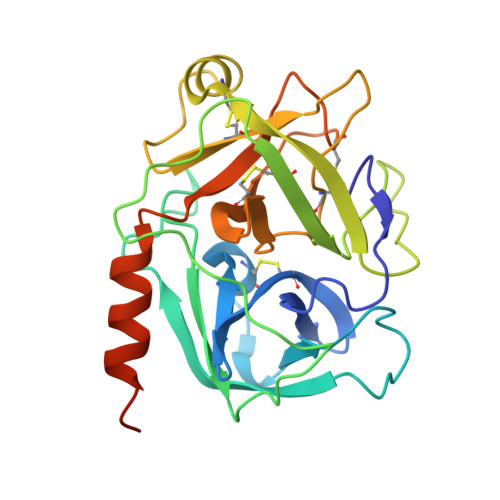
4Q7X

Neutrophil serine protease 4 (PRSS57) apo form 1


Experimental Data Snapshot

  • Method: X-RAY DIFFRACTION
  • Resolution: 2.55 Å
  • R-Value Free: 0.231 
  • R-Value Work: 0.197 
  • R-Value Observed: 0.199 

Starting Model: experimental
View more details

wwPDB Validation   3D Report Full Report


Ligand Structure Quality Assessment 


This is version 2.1 of the entry. See complete history


Literature

Structures of neutrophil serine protease 4 reveal an unusual mechanism of substrate recognition by a trypsin-fold protease.

Lin, S.J.Dong, K.C.Eigenbrot, C.van Lookeren Campagne, M.Kirchhofer, D.

(2014) Structure 22: 1333-1340

  • DOI: https://doi.org/10.1016/j.str.2014.07.008
  • Primary Citation of Related Structures:  
    4Q7X, 4Q7Y, 4Q7Z, 4Q80

  • PubMed Abstract: 

    Trypsin-fold proteases, the largest mammalian protease family, are classified by their primary substrate specificity into one of three categories, trypsin-like, chymotrypsin-like, and elastase-like, based on key structural features of their active site. However, the recently discovered neutrophil serine protease 4 (NSP4, also known as PRSS57) presents a paradox: NSP4 exhibits a trypsin-like specificity for cleaving substrates after arginine residues, but it bears elastase-like specificity determining residues in the active site. Here we show that NSP4 has a fully occluded S1 pocket and that the substrate P1-arginine adopts a noncanonical "up" conformation stabilized by a solvent-exposed H-bond network. This uncommon arrangement, conserved in all NSP4 orthologs, enables NSP4 to process substrates after both arginine as well as post-translationally modified arginine residues, such as methylarginine and citrulline. These findings establish a distinct paradigm for substrate recognition by a trypsin-fold protease and provide insights into the function of NSP4.


  • Organizational Affiliation

    Department of Early Discovery Biochemistry, Genentech, Inc., 1 DNA Way, South San Francisco, CA 94080, USA.


Macromolecules
Find similar proteins by:  (by identity cutoff)  |  3D Structure
Entity ID: 1
MoleculeChains Sequence LengthOrganismDetailsImage
Serine protease 57
A, B
250Homo sapiensMutation(s): 0 
Gene Names: PRSS57PRSSL1UNQ782/PRO1599
EC: 3.4.21
UniProt & NIH Common Fund Data Resources
Find proteins for Q6UWY2 (Homo sapiens)
Explore Q6UWY2 
Go to UniProtKB:  Q6UWY2
PHAROS:  Q6UWY2
GTEx:  ENSG00000185198 
Entity Groups  
Sequence Clusters30% Identity50% Identity70% Identity90% Identity95% Identity100% Identity
UniProt GroupQ6UWY2
Glycosylation
Glycosylation Sites: 2Go to GlyGen: Q6UWY2-1
Sequence Annotations
Expand
  • Reference Sequence
Oligosaccharides

Help

Entity ID: 2
MoleculeChains Length2D Diagram Glycosylation3D Interactions
alpha-L-fucopyranose-(1-6)-2-acetamido-2-deoxy-beta-D-glucopyranose
C, D
2N-Glycosylation
Glycosylation Resources
GlyTouCan:  G86851RC
GlyCosmos:  G86851RC
GlyGen:  G86851RC
Experimental Data & Validation

Experimental Data

  • Method: X-RAY DIFFRACTION
  • Resolution: 2.55 Å
  • R-Value Free: 0.231 
  • R-Value Work: 0.197 
  • R-Value Observed: 0.199 
  • Space Group: P 41
Unit Cell:
Length ( Å )Angle ( ˚ )
a = 70.366α = 90
b = 70.366β = 90
c = 150.021γ = 90
Software Package:
Software NamePurpose
BOSdata collection
BUSTERrefinement
HKL-2000data reduction
HKL-2000data scaling

Structure Validation

View Full Validation Report


Ligand Structure Quality Assessment 


Entry History 

Deposition Data

Revision History  (Full details and data files)

  • Version 1.0: 2014-09-03
    Type: Initial release
  • Version 1.1: 2014-09-17
    Changes: Database references
  • Version 1.2: 2017-11-22
    Changes: Refinement description
  • Version 2.0: 2020-07-29
    Type: Remediation
    Reason: Carbohydrate remediation
    Changes: Atomic model, Data collection, Derived calculations, Structure summary
  • Version 2.1: 2023-09-20
    Changes: Data collection, Database references, Refinement description, Structure summary