4Q5I | pdb_00004q5i

Crystal structure of a T151A mutant of the E. coli FeoB G-domain


Experimental Data Snapshot

  • Method: X-RAY DIFFRACTION
  • Resolution: 2.80 Å
  • R-Value Free: 
    0.298 (Depositor), 0.295 (DCC) 
  • R-Value Work: 
    0.239 (Depositor), 0.236 (DCC) 
  • R-Value Observed: 
    0.241 (Depositor) 

wwPDB Validation 3D Report Full Report

Validation slider image for 4Q5I

This is version 1.1 of the entry. See complete history

Literature

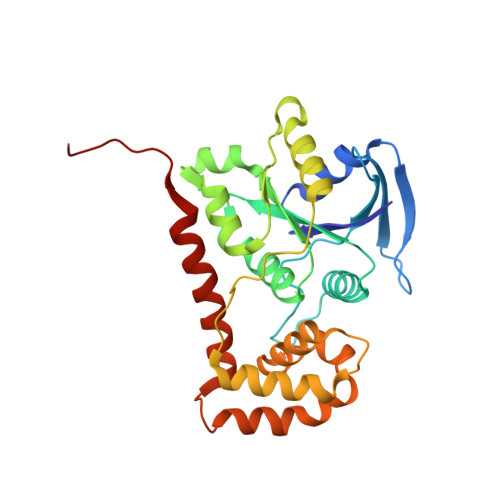
Exploring the correlation between the sequence composition of the nucleotide binding G5 loop of the FeoB GTPase domain (NFeoB) and intrinsic rate of GDP release.

Guilfoyle, A.P.Deshpande, C.N.Schenk, G.Maher, M.J.Jormakka, M.

(2014) Biosci Rep 34: e00158-e00158

  • DOI: https://doi.org/10.1042/BSR20140152
  • Primary Citation Related Structures: 
    4Q00, 4Q5I

  • PubMed Abstract: 

    GDP release from GTPases is usually extremely slow and is in general assisted by external factors, such as association with guanine exchange factors or membrane-embedded GPCRs (G protein-coupled receptors), which accelerate the release of GDP by several orders of magnitude. Intrinsic factors can also play a significant role; a single amino acid substitution in one of the guanine nucleotide recognition motifs, G5, results in a drastically altered GDP release rate, indicating that the sequence composition of this motif plays an important role in spontaneous GDP release. In the present study, we used the GTPase domain from EcNFeoB (Escherichia coli FeoB) as a model and applied biochemical and structural approaches to evaluate the role of all the individual residues in the G5 loop. Our study confirms that several of the residues in the G5 motif have an important role in the intrinsic affinity and release of GDP. In particular, a T151A mutant (third residue of the G5 loop) leads to a reduced nucleotide affinity and provokes a drastically accelerated dissociation of GDP.


  • Organizational Affiliation
    • *Structural Biology Program, Centenary Institute, Locked Bag 6, Sydney, New South Wales 2042, Australia.

Macromolecule Content 

  • Total Structure Weight: 178.2 kDa 
  • Atom Count: 12,130 
  • Modeled Residue Count: 1,584 
  • Deposited Residue Count: 1,620 
  • Unique protein chains: 1

Macromolecules

Find similar proteins by:|  3D Structure
Entity ID: 1
MoleculeChains  Sequence LengthOrganismDetailsImage
Ferrous iron transport protein B
A, B, C, D, E
A, B, C, D, E, F
270Escherichia coli K-12Mutation(s): 1 
Gene Names: feoBb3409JW3372
UniProt
Find proteins for P33650 (Escherichia coli (strain K12))
Explore P33650 
Go to UniProtKB:  P33650
Entity Groups
Sequence Clusters30% Identity50% Identity70% Identity90% Identity95% Identity100% Identity
UniProt GroupP33650
Sequence Annotations
Expand
Reference Sequence

Small Molecules

Ligands 1 Unique
IDChains Name / Formula / InChI Key2D Diagram3D Interactions
SO4

Query on SO4


Download:Ideal Coordinates CCD File
G [auth D]SULFATE ION
O4 S
QAOWNCQODCNURD-UHFFFAOYSA-L

Experimental Data & Validation

Experimental Data

  • Method: X-RAY DIFFRACTION
  • Resolution: 2.80 Å
  • R-Value Free:  0.298 (Depositor), 0.295 (DCC) 
  • R-Value Work:  0.239 (Depositor), 0.236 (DCC) 
  • R-Value Observed: 0.241 (Depositor) 
Space Group: P 1
Unit Cell:
Length ( Å )Angle ( ˚ )
a = 60.978α = 115.67
b = 82.779β = 108.5
c = 95.505γ = 90.06
Software Package:
Software NamePurpose
SCALEPACKdata scaling
REFMACrefinement
PDB_EXTRACTdata extraction
MARmosaicdata collection
MOSFLMdata reduction
PHASERphasing

Structure Validation

View Full Validation Report


Entry History 

Deposition Data

Revision History  (Full details and data files)

  • Version 1.0: 2015-04-08
    Type: Initial release
  • Version 1.1: 2024-03-20
    Changes: Data collection, Database references, Derived calculations