4Q0Z | pdb_00004q0z

The catalytic core of Rad2 in complex with DNA substrate (complex III)


Experimental Data Snapshot

  • Method: X-RAY DIFFRACTION
  • Resolution: 2.40 Å
  • R-Value Free: 
    0.235 (Depositor), 0.237 (DCC) 
  • R-Value Work: 
    0.178 (Depositor), 0.180 (DCC) 
  • R-Value Observed: 
    0.181 (Depositor) 

Starting Model: experimental
View more details

wwPDB Validation 3D Report Full Report

Validation slider image for 4Q0Z

This is version 1.5 of the entry. See complete history

Literature

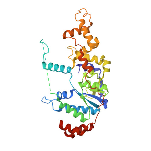
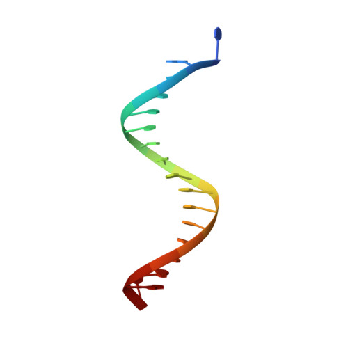
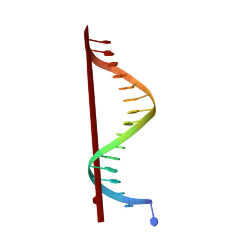
Crystal structure of the catalytic core of Rad2: insights into the mechanism of substrate binding.

Mietus, M.Nowak, E.Jaciuk, M.Kustosz, P.Studnicka, J.Nowotny, M.

(2014) Nucleic Acids Res 42: 10762-10775

  • DOI: https://doi.org/10.1093/nar/gku729
  • Primary Citation Related Structures: 
    4Q0R, 4Q0W, 4Q0Z, 4Q10

  • PubMed Abstract: 

    Rad2/XPG belongs to the flap nuclease family and is responsible for a key step of the eukaryotic nucleotide excision DNA repair (NER) pathway. To elucidate the mechanism of DNA binding by Rad2/XPG, we solved crystal structures of the catalytic core of Rad2 in complex with a substrate. Rad2 utilizes three structural modules for recognition of the double-stranded portion of DNA substrate, particularly a Rad2-specific α-helix for binding the cleaved strand. The protein does not specifically recognize the single-stranded portion of the nucleic acid. Our data suggest that in contrast to related enzymes (FEN1 and EXO1), the Rad2 active site may be more accessible, which would create an exit route for substrates without a free 5' end.


  • Organizational Affiliation
    • Laboratory of Protein Structure, International Institute of Molecular and Cell Biology, Warsaw 02-109, Poland.

Macromolecule Content 

  • Total Structure Weight: 189.87 kDa 
  • Atom Count: 13,433 
  • Modeled Residue Count: 1,308 
  • Deposited Residue Count: 1,528 
  • Unique protein chains: 1
  • Unique nucleic acid chains: 2

Macromolecules


Find similar proteins by:|  3D Structure
Entity ID: 1
MoleculeChains  Sequence LengthOrganismDetailsImage
Rad2p
A, B, E, F
365Saccharomyces cerevisiae S288CMutation(s): 0 
Gene Names: RAD2YGR258C
EC: 3.1
UniProt
Find proteins for P07276 (Saccharomyces cerevisiae (strain ATCC 204508 / S288c))
Explore P07276 
Go to UniProtKB:  P07276
Entity Groups
Sequence Clusters30% Identity50% Identity70% Identity90% Identity95% Identity100% Identity
UniProt GroupP07276
Sequence Annotations
Expand
Reference Sequence
Find similar nucleic acids by:  Sequence
Entity ID: 2
MoleculeChains LengthOrganismImage
DNA (5'-D(*TP*GP*CP*TP*CP*CP*CP*TP*TP*GP*TP*CP*TP*CP*AP*GP*T)-3')
C, G
17N/A
Sequence Annotations
Expand
Reference Sequence
Find similar nucleic acids by:  Sequence
Entity ID: 3
MoleculeChains LengthOrganismImage
DNA (5'-D(*TP*CP*TP*GP*AP*GP*AP*CP*AP*AP*GP*GP*GP*AP*GP*CP*T)-3')
D, H
17N/A
Sequence Annotations
Expand
Reference Sequence

Small Molecules

Ligands 2 Unique
IDChains Name / Formula / InChI Key2D Diagram3D Interactions
CA

Query on CA


Download:Ideal Coordinates CCD File
I [auth A],
K [auth B],
M [auth E],
O [auth F]
CALCIUM ION
Ca
BHPQYMZQTOCNFJ-UHFFFAOYSA-N
K

Query on K


Download:Ideal Coordinates CCD File
J [auth A],
L [auth B],
N [auth E],
P [auth F]
POTASSIUM ION
K
NPYPAHLBTDXSSS-UHFFFAOYSA-N

Experimental Data & Validation

Experimental Data

  • Method: X-RAY DIFFRACTION
  • Resolution: 2.40 Å
  • R-Value Free:  0.235 (Depositor), 0.237 (DCC) 
  • R-Value Work:  0.178 (Depositor), 0.180 (DCC) 
  • R-Value Observed: 0.181 (Depositor) 
Space Group: P 1 21 1
Unit Cell:
Length ( Å )Angle ( ˚ )
a = 110.278α = 90
b = 86.039β = 110.95
c = 134.44γ = 90
Software Package:
Software NamePurpose
MAR345data collection
PHASERphasing
PHENIXrefinement
HKL-2000data reduction
HKL-2000data scaling

Structure Validation

View Full Validation Report


Entry History 

Deposition Data

Revision History  (Full details and data files)

  • Version 1.0: 2014-08-27
    Type: Initial release
  • Version 1.1: 2014-09-17
    Changes: Structure summary
  • Version 1.2: 2014-10-01
    Changes: Database references
  • Version 1.3: 2017-08-09
    Changes: Refinement description, Source and taxonomy
  • Version 1.4: 2017-11-22
    Changes: Refinement description
  • Version 1.5: 2023-09-20
    Changes: Data collection, Database references, Derived calculations, Refinement description