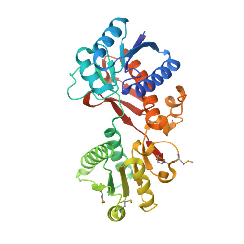
Structure of a putative branched-chain amino acid ABC transporter from Chromobacterium violaceum ATCC 12472
Filippova, E.V., Minasov, G., Shuvalova, L., Kiryukhina, O., Endres, M., Joachimiak, A., Anderson, W.F., Midwest Center for Structural Genomics (MCSG)To be published.