Structural and Spectroscopic Insights into BolA-Glutaredoxin Complexes.
Roret, T., Tsan, P., Couturier, J., Zhang, B., Johnson, M.K., Rouhier, N., Didierjean, C.(2014) J Biol Chem 289: 24588-24598
- PubMed: 25012657
- DOI: https://doi.org/10.1074/jbc.M114.572701
- Primary Citation of Related Structures:
2MM9, 2MMA, 4PUG, 4PUH, 4PUI - PubMed Abstract:
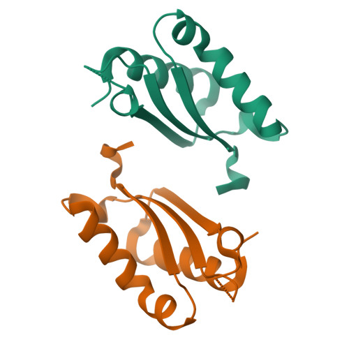
BolA proteins are defined as stress-responsive transcriptional regulators, but they also participate in iron metabolism. Although they can form [2Fe-2S]-containing complexes with monothiol glutaredoxins (Grx), structural details are lacking. Three Arabidopsis thaliana BolA structures were solved. They differ primarily by the size of a loop referred to as the variable [H/C] loop, which contains an important cysteine (BolA_C group) or histidine (BolA_H group) residue. From three-dimensional modeling and spectroscopic analyses of A. thaliana GrxS14-BolA1 holo-heterodimer (BolA_H), we provide evidence for the coordination of a Rieske-type [2Fe-2S] cluster. For BolA_C members, the cysteine could replace the histidine as a ligand. NMR interaction experiments using apoproteins indicate that a completely different heterodimer was formed involving the nucleic acid binding site of BolA and the C-terminal tail of Grx. The possible biological importance of these complexes is discussed considering the physiological functions previously assigned to BolA and to Grx-BolA or Grx-Grx complexes.
Organizational Affiliation:
From the Université de Lorraine and CNRS, UMR 7036 CRM2, BioMod group, 54506 Vandœuvre-lès-Nancy, France.


