4PU0

Crystal structure of the Escherichia coli alkanesulfonate FMN reductase SsuE in FMNH2-bound form


Experimental Data Snapshot

  • Method: X-RAY DIFFRACTION
  • Resolution: 2.30 Å
  • R-Value Free: 0.221 
  • R-Value Work: 0.173 
  • R-Value Observed: 0.176 

Starting Model: experimental
View more details

wwPDB Validation   3D Report Full Report


Ligand Structure Quality Assessment 


This is version 1.2 of the entry. See complete history


Literature

Crystal Structure of Escherichia coli SsuE: Defining a General Catalytic Cycle for FMN Reductases of the Flavodoxin-like Superfamily.

Driggers, C.M.Dayal, P.V.Ellis, H.R.Karplus, P.A.

(2014) Biochemistry 53: 3509-3519

  • DOI: https://doi.org/10.1021/bi500314f
  • Primary Citation of Related Structures:  
    4PTY, 4PTZ, 4PU0

  • PubMed Abstract: 

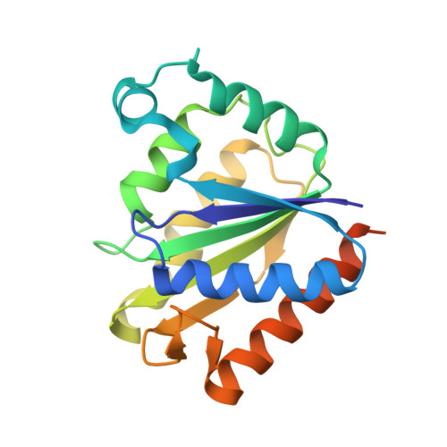
    The Escherichia coli sulfur starvation utilization (ssu) operon includes a two-component monooxygenase system consisting of a nicotinamide adenine dinucleotide phosphate (NADPH)-dependent flavin mononucleotide (FMN) reductase, SsuE, and a monooxygenase, SsuD. SsuE is part of the flavodoxin-like superfamily, and we report here the crystal structures of its apo, FMN-bound, and FMNH2-bound forms at ∼2 Å resolution. In the crystals, SsuE forms a tetramer that is a dimer of dimers similar to those seen for homologous FMN reductases, quinone reductases, and the WrbA family of enzymes. A π-helix present at the tetramer building interface is unique to the reductases from two-component monooxygenase systems. Analytical ultracentrifugation studies of SsuE confirm a dimer-tetramer equilibrium exists in solution, with FMN binding favoring the dimer. As the active site includes residues from both subunits, at least a dimeric association is required for the function of SsuE. The structures show that one FMN binds tightly in a deeply held site, which makes available a second binding site, in which either a second FMN or the nicotinamide of NADPH can bind. The FMNH2-bound structure shows subtle changes consistent with its binding being weaker than that of FMN. Combining this information with published kinetic studies, we propose a general catalytic cycle for two-component reductases of the flavodoxin-like superfamily, by which the enzyme can potentially provide FMNH2 to its partner monooxygenase by different routes depending on the FMN concentration and the presence of a partner monooxygenase.


  • Organizational Affiliation

    Department of Biochemistry and Biophysics, Oregon State University , 2011 Agricultural and Life Sciences Building, Corvallis, Oregon 97331, United States.


Macromolecules
Find similar proteins by:  (by identity cutoff)  |  3D Structure
Entity ID: 1
MoleculeChains Sequence LengthOrganismDetailsImage
FMN reductase SsuEA [auth C],
B [auth A],
C [auth B],
D
191Escherichia coli K-12Mutation(s): 0 
Gene Names: ssuEycbPb0937JW0920
EC: 1.5.1.38
UniProt
Find proteins for P80644 (Escherichia coli (strain K12))
Explore P80644 
Go to UniProtKB:  P80644
Entity Groups  
Sequence Clusters30% Identity50% Identity70% Identity90% Identity95% Identity100% Identity
UniProt GroupP80644
Sequence Annotations
Expand
  • Reference Sequence
Small Molecules
Ligands 3 Unique
IDChains Name / Formula / InChI Key2D Diagram3D Interactions
FMN
Query on FMN

Download Ideal Coordinates CCD File 
E [auth C],
G [auth A],
J [auth D]
FLAVIN MONONUCLEOTIDE
C17 H21 N4 O9 P
FVTCRASFADXXNN-SCRDCRAPSA-N
PO4
Query on PO4

Download Ideal Coordinates CCD File 
H [auth A],
I [auth B]
PHOSPHATE ION
O4 P
NBIIXXVUZAFLBC-UHFFFAOYSA-K
GOL
Query on GOL

Download Ideal Coordinates CCD File 
F [auth C],
K [auth D],
L [auth D]
GLYCEROL
C3 H8 O3
PEDCQBHIVMGVHV-UHFFFAOYSA-N
Experimental Data & Validation

Experimental Data

  • Method: X-RAY DIFFRACTION
  • Resolution: 2.30 Å
  • R-Value Free: 0.221 
  • R-Value Work: 0.173 
  • R-Value Observed: 0.176 
  • Space Group: P 61 2 2
Unit Cell:
Length ( Å )Angle ( ˚ )
a = 187.018α = 90
b = 187.018β = 90
c = 91.336γ = 120
Software Package:
Software NamePurpose
ADSCdata collection
PHASERphasing
PHENIXrefinement
HKL-2000data reduction
HKL-2000data scaling

Structure Validation

View Full Validation Report


Ligand Structure Quality Assessment 


Entry History 

Deposition Data

Revision History  (Full details and data files)

  • Version 1.0: 2014-06-18
    Type: Initial release
  • Version 1.1: 2024-02-28
    Changes: Data collection, Database references, Derived calculations
  • Version 1.2: 2024-04-03
    Changes: Refinement description