4PSO | pdb_00004pso

Crystal structure of apeThermo-DBP-RP2 bound to ssDNA dT10


Experimental Data Snapshot

  • Method: X-RAY DIFFRACTION
  • Resolution: 2.90 Å
  • R-Value Free: 
    0.309 (Depositor), 0.311 (DCC) 
  • R-Value Work: 
    0.267 (Depositor), 0.270 (DCC) 
  • R-Value Observed: 
    0.269 (Depositor) 

Starting Model: experimental
View more details

wwPDB Validation 3D Report Full Report

Validation slider image for 4PSO

This is version 1.2 of the entry. See complete history

Literature

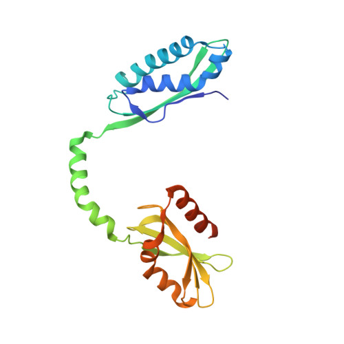
Entrapment of DNA in an intersubunit tunnel system of a single-stranded DNA-binding protein.

Ghalei, H.Moeller, H.v.Eppers, D.Sohmen, D.Wilson, D.N.Loll, B.Wahl, M.C.

(2014) Nucleic Acids Res 42: 6698-6708

  • DOI: https://doi.org/10.1093/nar/gku259
  • Primary Citation Related Structures: 
    4PSL, 4PSM, 4PSN, 4PSO

  • PubMed Abstract: 

    Instead of a classical single-stranded deoxyribonuleic acid (DNA)-binding protein (SSB), some hyperthermophilic crenarchaea harbor a non-canonical SSB termed ThermoDBP. Two related but poorly characterized groups of proteins, which share the ThermoDBP N-terminal DNA-binding domain, have a broader phylogenetic distribution and co-exist with ThermoDBPs and/or other SSBs. We have investigated the nucleic acid binding properties and crystal structures of representatives of these groups of ThermoDBP-related proteins (ThermoDBP-RPs) 1 and 2. ThermoDBP-RP 1 and 2 oligomerize by different mechanisms and only ThermoDBP-RP2 exhibits strong single-stranded DNA affinity in vitro. A crystal structure of ThermoDBP-RP2 in complex with DNA reveals how the NTD common to ThermoDBPs and ThermoDBP-RPs can contact the nucleic acid in a manner that allows a symmetric homotetrameric protein complex to bind single-stranded DNA molecules asymmetrically. While single-stranded DNA wraps around the surface or binds along channels of previously investigated SSBs, it traverses an internal, intersubunit tunnel system of a ThermoDBP-RP2 tetramer. Our results indicate that some archaea have acquired special SSBs for genome maintenance in particularly challenging environments.


  • Organizational Affiliation
    • Freie Universität Berlin, Fachbereich Biologie, Chemie, Pharmazie, Institut für Chemie und Biochemie, AG Strukturbiochemie, Takustr. 6, 14195 Berlin, Germany Max-Planck-Institute for Biophysical Chemistry, Department of Cellular Biochemistry, Am Fassberg 11, 37077 Göttingen, Germany.

Macromolecule Content 

  • Total Structure Weight: 225.1 kDa 
  • Atom Count: 14,476 
  • Modeled Residue Count: 1,779 
  • Deposited Residue Count: 1,936 
  • Unique protein chains: 1
  • Unique nucleic acid chains: 1

Macromolecules


Find similar proteins by:|  3D Structure
Entity ID: 1
MoleculeChains  Sequence LengthOrganismDetailsImage
ssDNA binding protein237Aeropyrum pernix K1Mutation(s): 0 
Gene Names: APE_1866.1
UniProt
Find proteins for Q9YAS7 (Aeropyrum pernix (strain ATCC 700893 / DSM 11879 / JCM 9820 / NBRC 100138 / K1))
Explore Q9YAS7 
Go to UniProtKB:  Q9YAS7
Entity Groups
Sequence Clusters30% Identity50% Identity70% Identity90% Identity95% Identity100% Identity
UniProt GroupQ9YAS7
Sequence Annotations
Expand
Reference Sequence
Find similar nucleic acids by:  Sequence
Entity ID: 2
MoleculeChains LengthOrganismImage
polydeoxyribonucleotideI [auth L],
J [auth E],
K [auth X],
L [auth Z]
10N/A
Sequence Annotations
Expand
Reference Sequence

Small Molecules

Ligands 1 Unique
IDChains Name / Formula / InChI Key2D Diagram3D Interactions
PO4

Query on PO4


Download:Ideal Coordinates CCD File
M [auth B]PHOSPHATE ION
O4 P
NBIIXXVUZAFLBC-UHFFFAOYSA-K

Experimental Data & Validation

Experimental Data

  • Method: X-RAY DIFFRACTION
  • Resolution: 2.90 Å
  • R-Value Free:  0.309 (Depositor), 0.311 (DCC) 
  • R-Value Work:  0.267 (Depositor), 0.270 (DCC) 
  • R-Value Observed: 0.269 (Depositor) 
Space Group: P 21 21 21
Unit Cell:
Length ( Å )Angle ( ˚ )
a = 120.488α = 90
b = 190.277β = 90
c = 89.786γ = 90
Software Package:
Software NamePurpose
MAR345dtbdata collection
PHASERphasing
PHENIXrefinement
XDSdata reduction
XSCALEdata scaling

Structure Validation

View Full Validation Report


Entry History 

Deposition Data

Revision History  (Full details and data files)

  • Version 1.0: 2014-04-30
    Type: Initial release
  • Version 1.1: 2014-06-25
    Changes: Database references
  • Version 1.2: 2023-09-20
    Changes: Data collection, Database references, Derived calculations, Refinement description