4PPJ | pdb_00004ppj

Crystal structure of Phanta, a weakly fluorescent photochromic GFP-like protein. ON state


Experimental Data Snapshot

  • Method: X-RAY DIFFRACTION
  • Resolution: 2.30 Å
  • R-Value Free: 
    0.263 (Depositor), 0.269 (DCC) 
  • R-Value Work: 
    0.197 (Depositor) 
  • R-Value Observed: 
    0.200 (Depositor) 

Starting Model: experimental
View more details

wwPDB Validation 3D Report Full Report

Validation slider image for 4PPJ

This is version 1.2 of the entry. See complete history

Literature

X-Ray Crystal Structure and Properties of Phanta, a Weakly Fluorescent Photochromic GFP-Like Protein.

Don Paul, C.Traore, D.A.Olsen, S.Devenish, R.J.Close, D.W.Bell, T.D.Bradbury, A.Wilce, M.C.Prescott, M.

(2015) PLoS One 10: e0123338-e0123338

  • DOI: https://doi.org/10.1371/journal.pone.0123338
  • Primary Citation Related Structures: 
    4PPJ, 4PPK, 4PPL

  • PubMed Abstract: 

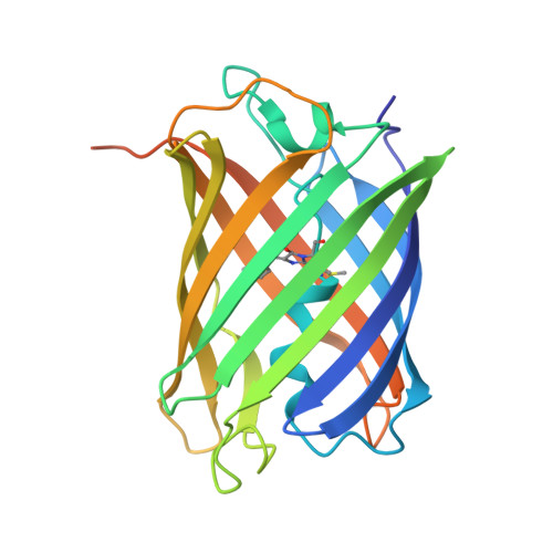
    Phanta is a reversibly photoswitching chromoprotein (ΦF, 0.003), useful for pcFRET, that was isolated from a mutagenesis screen of the bright green fluorescent eCGP123 (ΦF, 0.8). We have investigated the contribution of substitutions at positions His193, Thr69 and Gln62, individually and in combination, to the optical properties of Phanta. Single amino acid substitutions at position 193 resulted in proteins with very low ΦF, indicating the importance of this position in controlling the fluorescence efficiency of the variant proteins. The substitution Thr69Val in Phanta was important for supressing the formation of a protonated chromophore species observed in some His193 substituted variants, whereas the substitution Gln62Met did not significantly contribute to the useful optical properties of Phanta. X-ray crystal structures for Phanta (2.3 Å), eCGP123T69V (2.0 Å) and eCGP123H193Q (2.2 Å) in their non-photoswitched state were determined, revealing the presence of a cis-coplanar chromophore. We conclude that changes in the hydrogen-bonding network supporting the cis-chromophore, and its contacts with the surrounding protein matrix, are responsible for the low fluorescence emission of eCGP123 variants containing a His193 substitution.


  • Organizational Affiliation
    • Department of Neuro- and Sensory Physiology, University Medicine, Göttingen, 37073, Göttingen, Germany.

Macromolecule Content 

  • Total Structure Weight: 112.99 kDa 
  • Atom Count: 7,291 
  • Modeled Residue Count: 865 
  • Deposited Residue Count: 980 
  • Unique protein chains: 1

Macromolecules

Find similar proteins by:|  3D Structure
Entity ID: 1
MoleculeChains  Sequence LengthOrganismDetailsImage
Monomeric Azami Green
A, B, C, D
245synthetic constructMutation(s): 0 

Small Molecules

Modified Residues  1 Unique
IDChains TypeFormula2D DiagramParent
NRQ
Query on NRQ
A, B, C, D
L-PEPTIDE LINKINGC16 H17 N3 O4 SMET, TYR, GLY

Experimental Data & Validation

Experimental Data

  • Method: X-RAY DIFFRACTION
  • Resolution: 2.30 Å
  • R-Value Free:  0.263 (Depositor), 0.269 (DCC) 
  • R-Value Work:  0.197 (Depositor) 
  • R-Value Observed: 0.200 (Depositor) 
Space Group: P 1 21 1
Unit Cell:
Length ( Å )Angle ( ˚ )
a = 69.815α = 90
b = 79.37β = 102.4
c = 71.001γ = 90
Software Package:
Software NamePurpose
Blu-Icedata collection
PHASERphasing
REFMACrefinement
XDSdata reduction
XSCALEdata scaling
BUSTERrefinement

Structure Validation

View Full Validation Report


Entry History 

Deposition Data

Revision History  (Full details and data files)

  • Version 1.0: 2015-04-08
    Type: Initial release
  • Version 1.1: 2015-05-20
    Changes: Database references
  • Version 1.2: 2024-04-03
    Changes: Data collection, Database references, Derived calculations, Refinement description