4POS

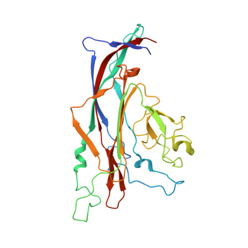
Structure of Human Polyomavirus 9 VP1 pentamer in complex with 3'-sialyllactosamine


Experimental Data Snapshot

  • Method: X-RAY DIFFRACTION
  • Resolution: 2.00 Å
  • R-Value Free: 0.213 
  • R-Value Work: 0.179 
  • R-Value Observed: 0.180 

Starting Model: experimental
View more details

wwPDB Validation   3D Report Full Report


This is version 2.2 of the entry. See complete history


Literature

Crystallographic and glycan microarray analysis of human polyomavirus 9 VP1 identifies N-glycolyl neuraminic acid as a receptor candidate.

Khan, Z.M.Liu, Y.Neu, U.Gilbert, M.Ehlers, B.Feizi, T.Stehle, T.

(2014) J Virol 88: 6100-6111

  • DOI: https://doi.org/10.1128/JVI.03455-13
  • Primary Citation of Related Structures:  
    4POQ, 4POR, 4POS, 4POT

  • PubMed Abstract: 

    Human polyomavirus 9 (HPyV9) is a closely related homologue of simian B-lymphotropic polyomavirus (LPyV). In order to define the architecture and receptor binding properties of HPyV9, we solved high-resolution crystal structures of its major capsid protein, VP1, in complex with three putative oligosaccharide receptors identified by glycan microarray screening. Comparison of the properties of HPyV9 VP1 with the known structure and glycan-binding properties of LPyV VP1 revealed that both viruses engage short sialylated oligosaccharides, but small yet important differences in specificity were detected. Surprisingly, HPyV9 VP1 preferentially binds sialyllactosamine compounds terminating in 5-N-glycolyl neuraminic acid (Neu5Gc) over those terminating in 5-N-acetyl neuraminic acid (Neu5Ac), whereas LPyV does not exhibit such a preference. The structural analysis demonstrated that HPyV9 makes specific contacts, via hydrogen bonds, with the extra hydroxyl group present in Neu5Gc. An equivalent hydrogen bond cannot be formed by LPyV VP1. The most common sialic acid in humans is 5-N-acetyl neuraminic acid (Neu5Ac), but various modifications give rise to more than 50 different sialic acid variants that decorate the cell surface. Unlike most mammals, humans cannot synthesize the sialic acid variant 5-N-glycolyl neuraminic acid (Neu5Gc) due to a gene defect. Humans can, however, still acquire this compound from dietary sources. The role of Neu5Gc in receptor engagement and in defining viral tropism is only beginning to emerge, and structural analyses defining the differences in specificity for Neu5Ac and Neu5Gc are still rare. Using glycan microarray screening and high-resolution protein crystallography, we have examined the receptor specificity of a recently discovered human polyomavirus, HPyV9, and compared it to that of the closely related simian polyomavirus LPyV. Our study highlights critical differences in the specificities of both viruses, contributing to an enhanced understanding of the principles that underlie pathogen selectivity for modified sialic acids.


  • Organizational Affiliation

    Interfaculty Institute of Biochemistry, University of Tuebingen, Tuebingen, Germany.


Macromolecules
Find similar proteins by:  (by identity cutoff)  |  3D Structure
Entity ID: 1
MoleculeChains Sequence LengthOrganismDetailsImage
VP1
A, B, C, D, E
A, B, C, D, E, F, G, H, I, J
278Human polyomavirus 9Mutation(s): 0 
Gene Names: gp3vp1
UniProt
Find proteins for E9NQ90 (Human polyomavirus 9)
Explore E9NQ90 
Go to UniProtKB:  E9NQ90
Entity Groups  
Sequence Clusters30% Identity50% Identity70% Identity90% Identity95% Identity100% Identity
UniProt GroupE9NQ90
Sequence Annotations
Expand
  • Reference Sequence
Oligosaccharides

Help

Entity ID: 2
MoleculeChains Length2D Diagram Glycosylation3D Interactions
N-acetyl-alpha-neuraminic acid-(2-3)-beta-D-galactopyranose-(1-4)-2-acetamido-2-deoxy-beta-D-glucopyranose
K, L, M, N, O
K, L, M, N, O, P, Q, R, S
3N/A
Glycosylation Resources
GlyTouCan:  G00065MO
GlyCosmos:  G00065MO
GlyGen:  G00065MO
Small Molecules
Ligands 3 Unique
IDChains Name / Formula / InChI Key2D Diagram3D Interactions
EDO
Query on EDO

Download Ideal Coordinates CCD File 
AB [auth G]
CA [auth B]
DA [auth B]
DB [auth H]
EA [auth B]
AB [auth G],
CA [auth B],
DA [auth B],
DB [auth H],
EA [auth B],
EB [auth H],
FA [auth B],
FB [auth H],
GB [auth H],
IA [auth C],
JB [auth I],
KB [auth I],
LA [auth D],
LB [auth I],
MA [auth D],
MB [auth I],
PA [auth E],
PB [auth J],
QA [auth E],
TA [auth F],
U [auth A],
UA [auth F],
V [auth A],
VA [auth F],
W [auth A],
WA [auth F],
X [auth A],
Y [auth A],
Z [auth A],
ZA [auth G]
1,2-ETHANEDIOL
C2 H6 O2
LYCAIKOWRPUZTN-UHFFFAOYSA-N
IPA
Query on IPA

Download Ideal Coordinates CCD File 
AA [auth A]
BB [auth G]
GA [auth B]
HB [auth H]
JA [auth C]
AA [auth A],
BB [auth G],
GA [auth B],
HB [auth H],
JA [auth C],
NA [auth D],
NB [auth I],
QB [auth J],
RA [auth E],
XA [auth F]
ISOPROPYL ALCOHOL
C3 H8 O
KFZMGEQAYNKOFK-UHFFFAOYSA-N
CA
Query on CA

Download Ideal Coordinates CCD File 
BA [auth B]
CB [auth H]
HA [auth C]
IB [auth I]
KA [auth D]
BA [auth B],
CB [auth H],
HA [auth C],
IB [auth I],
KA [auth D],
OA [auth E],
OB [auth J],
SA [auth F],
T [auth A],
YA [auth G]
CALCIUM ION
Ca
BHPQYMZQTOCNFJ-UHFFFAOYSA-N
Experimental Data & Validation

Experimental Data

  • Method: X-RAY DIFFRACTION
  • Resolution: 2.00 Å
  • R-Value Free: 0.213 
  • R-Value Work: 0.179 
  • R-Value Observed: 0.180 
  • Space Group: P 21 21 21
Unit Cell:
Length ( Å )Angle ( ˚ )
a = 96.91α = 90
b = 180.63β = 90
c = 199.42γ = 90
Software Package:
Software NamePurpose
RemDAqdata collection
MOLREPphasing
REFMACrefinement
XDSdata reduction
XSCALEdata scaling

Structure Validation

View Full Validation Report


Entry History 

Deposition Data

Revision History  (Full details and data files)

  • Version 1.0: 2014-04-09
    Type: Initial release
  • Version 1.1: 2014-11-26
    Changes: Database references
  • Version 2.0: 2020-07-29
    Type: Remediation
    Reason: Carbohydrate remediation
    Changes: Advisory, Atomic model, Data collection, Database references, Derived calculations, Structure summary
  • Version 2.1: 2023-09-20
    Changes: Advisory, Data collection, Database references, Refinement description, Structure summary
  • Version 2.2: 2024-11-20
    Changes: Structure summary