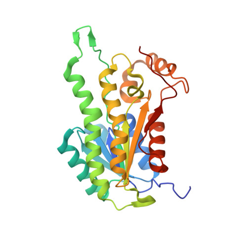
Crystal structure of 3-hydroxyacyl-CoA-dehydrogenase from Brucella melitensis
Lukacs, C.M., Abendroth, J., Edwards, T.E., Lorimer, D.To be published.
Experimental Data Snapshot
Starting Model: experimental
View more details
wwPDB Validation 3D Report Full Report

| Ligands 1 Unique | |||||
|---|---|---|---|---|---|
| ID | Chains | Name / Formula / InChI Key | 2D Diagram | 3D Interactions | |
| TRS Download:Ideal Coordinates CCD File | I [auth B], J [auth D], K [auth F], L [auth G] | 2-AMINO-2-HYDROXYMETHYL-PROPANE-1,3-DIOL C4 H12 N O3 LENZDBCJOHFCAS-UHFFFAOYSA-O |  | ||
| Length ( Å ) | Angle ( ˚ ) |
|---|---|
| a = 72.84 | α = 90 |
| b = 72.84 | β = 90 |
| c = 350.41 | γ = 90 |
| Software Name | Purpose |
|---|---|
| REFMAC | refinement |