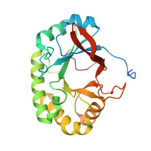
4PMO

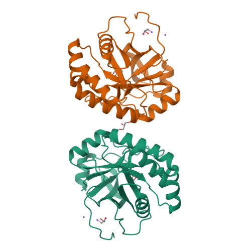
Crystal structure of the Mycobacterium tuberculosis Tat-secreted protein Rv2525c, monoclinic crystal form I


Experimental Data Snapshot

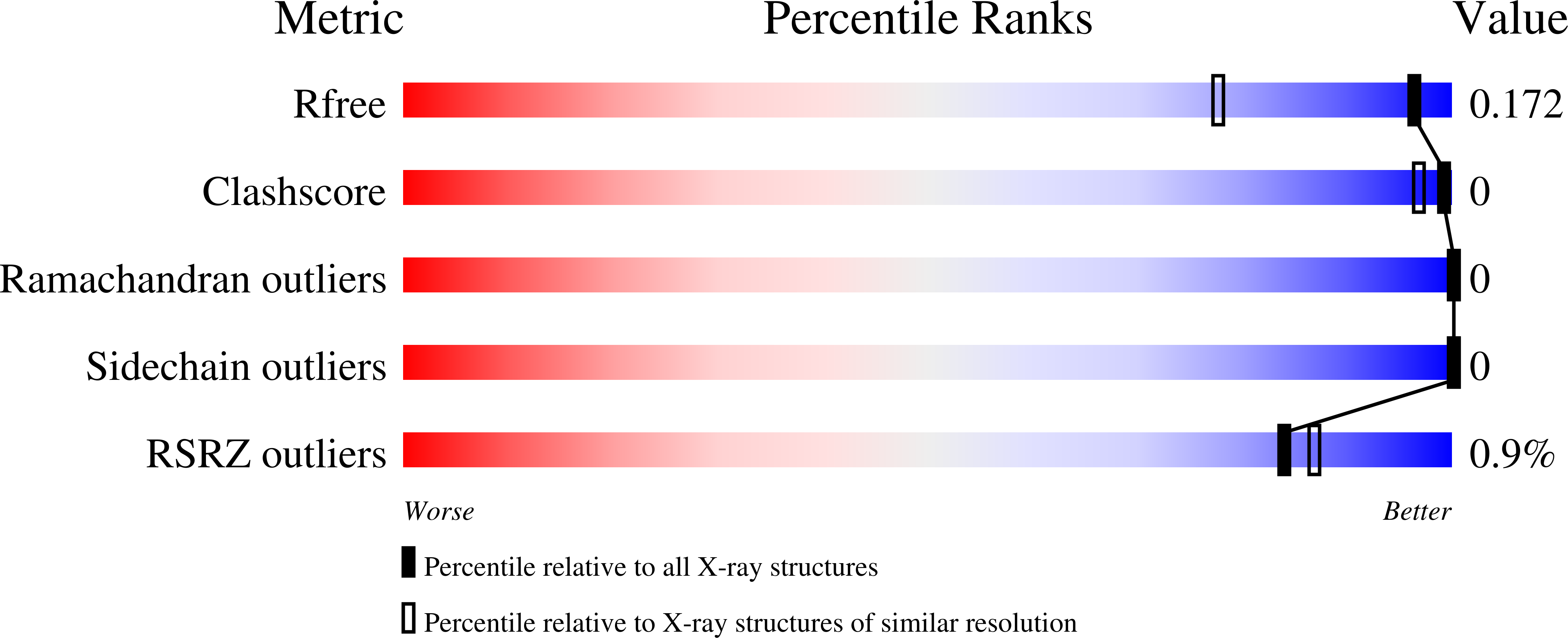
  • Method: X-RAY DIFFRACTION
  • Resolution: 1.33 Å
  • R-Value Free: 0.165 
  • R-Value Work: 0.149 
  • R-Value Observed: 0.150 

Starting Model: experimental
View more details

wwPDB Validation   3D Report Full Report


This is version 1.2 of the entry. See complete history


Literature

Structural studies suggest a peptidoglycan hydrolase function for the Mycobacterium tuberculosis Tat-secreted protein Rv2525c.

Bellinzoni, M.Haouz, A.Miras, I.Magnet, S.Andre-Leroux, G.Mukherjee, R.Shepard, W.Cole, S.T.Alzari, P.M.

(2014) J Struct Biol 188: 156-164

  • DOI: https://doi.org/10.1016/j.jsb.2014.09.003
  • Primary Citation of Related Structures:  
    4PMN, 4PMO, 4PMQ, 4PMR

  • PubMed Abstract: 

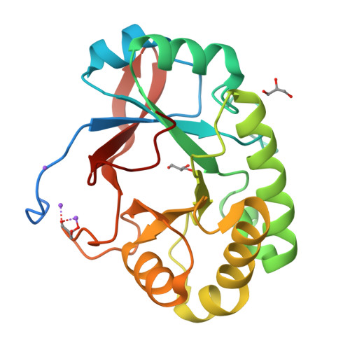
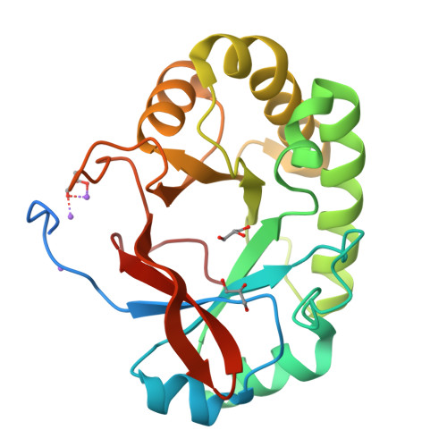
    Among the few proteins shown to be secreted by the Tat system in Mycobacterium tuberculosis, Rv2525c is of particular interest, since its gene is conserved in the minimal genome of Mycobacterium leprae. Previous evidence linked this protein to cell wall metabolism and sensitivity to β-lactams. We describe here the crystal structure of Rv2525c that shows a TIM barrel-like fold characteristic of glycoside hydrolases of the GH25 family, which includes prokaryotic and phage-encoded peptidoglycan hydrolases. Structural comparison with other members of this family combined with substrate docking suggest that, although the 'neighbouring group' catalytic mechanism proposed for this family still appears as the most plausible, the identity of residues involved in catalysis in GH25 hydrolases might need to be revised.


  • Organizational Affiliation

    Institut Pasteur, Unité de Microbiologie Structurale and CNRS-UMR3528, 25 rue du Dr. Roux, 75724 Paris Cedex 15, France. Electronic address: marco.bellinzoni@pasteur.fr.


Macromolecules
Find similar proteins by:  (by identity cutoff)  |  3D Structure
Entity ID: 1
MoleculeChains Sequence LengthOrganismDetailsImage
Tat-secreted protein Rv2525c
A, B
234Mycobacterium tuberculosis CDC1551Mutation(s): 0 
Gene Names: MT2601
UniProt
Find proteins for P95028 (Mycobacterium tuberculosis (strain CDC 1551 / Oshkosh))
Explore P95028 
Go to UniProtKB:  P95028
Entity Groups  
Sequence Clusters30% Identity50% Identity70% Identity90% Identity95% Identity100% Identity
UniProt GroupP95028
Sequence Annotations
Expand
  • Reference Sequence
Small Molecules
Ligands 3 Unique
IDChains Name / Formula / InChI Key2D Diagram3D Interactions
GOL
Query on GOL

Download Ideal Coordinates CCD File 
C [auth A]
D [auth A]
E [auth A]
O [auth B]
P [auth B]
C [auth A],
D [auth A],
E [auth A],
O [auth B],
P [auth B],
Q [auth B]
GLYCEROL
C3 H8 O3
PEDCQBHIVMGVHV-UHFFFAOYSA-N
FMT
Query on FMT

Download Ideal Coordinates CCD File 
F [auth A]
G [auth A]
H [auth A]
I [auth A]
J [auth A]
F [auth A],
G [auth A],
H [auth A],
I [auth A],
J [auth A],
K [auth A],
R [auth B],
S [auth B],
T [auth B],
U [auth B]
FORMIC ACID
C H2 O2
BDAGIHXWWSANSR-UHFFFAOYSA-N
NA
Query on NA

Download Ideal Coordinates CCD File 
L [auth A]
M [auth A]
N [auth A]
V [auth B]
W [auth B]
L [auth A],
M [auth A],
N [auth A],
V [auth B],
W [auth B],
X [auth B]
SODIUM ION
Na
FKNQFGJONOIPTF-UHFFFAOYSA-N
Experimental Data & Validation

Experimental Data

  • Method: X-RAY DIFFRACTION
  • Resolution: 1.33 Å
  • R-Value Free: 0.165 
  • R-Value Work: 0.149 
  • R-Value Observed: 0.150 
  • Space Group: C 1 2 1
Unit Cell:
Length ( Å )Angle ( ˚ )
a = 99.442α = 90
b = 73.679β = 130.85
c = 74.415γ = 90
Software Package:
Software NamePurpose
REFMACrefinement
XDSdata reduction
Aimlessdata scaling
PHASERphasing

Structure Validation

View Full Validation Report


Entry History 

Deposition Data

Revision History  (Full details and data files)

  • Version 1.0: 2014-10-08
    Type: Initial release
  • Version 1.1: 2014-12-10
    Changes: Database references
  • Version 1.2: 2023-12-20
    Changes: Data collection, Database references, Derived calculations, Refinement description