4PMJ | pdb_00004pmj

Crystal structure of a putative oxidoreductase from Sinorhizobium meliloti 1021 in complex with NADP


Experimental Data Snapshot

  • Method: X-RAY DIFFRACTION
  • Resolution: 2.20 Å
  • R-Value Free: 
    0.217 (Depositor), 0.225 (DCC) 
  • R-Value Work: 
    0.167 (Depositor), 0.182 (DCC) 
  • R-Value Observed: 
    0.170 (Depositor) 

wwPDB Validation 3D Report Full Report

Validation slider image for 4PMJ

Ligand Structure Quality Assessment 


This is version 1.6 of the entry. See complete history

Literature

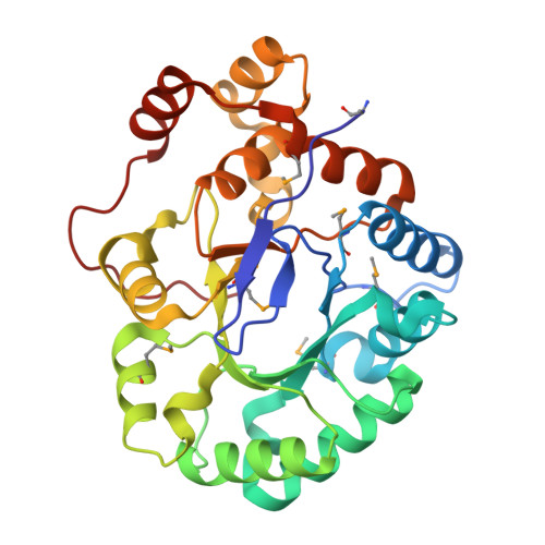
Crystal structure of a putative oxidoreductase from Sinorhizobiummeliloti 1021 in complex with NADP

Gasiorowska, O.A.Shabalin, I.G.Handing, K.B.Szlachta, K.Zimmerman, M.D.Hillerich, B.S.Gizzi, A.Toro, R.Bonanno, J.Seidel, R.Almo, S.C.Minor, W.

To be published.

Macromolecule Content 

  • Total Structure Weight: 31.92 kDa 
  • Atom Count: 2,305 
  • Modeled Residue Count: 281 
  • Deposited Residue Count: 281 
  • Unique protein chains: 1

Macromolecules

Find similar proteins by:|  3D Structure
Entity ID: 1
MoleculeChains  Sequence LengthOrganismDetailsImage
Putative oxidoreductase281Sinorhizobium meliloti 1021Mutation(s): 0 
Gene Names: R02112SMc01429
EC: 1.1.1
UniProt
Find proteins for Q92NR7 (Rhizobium meliloti (strain 1021))
Explore Q92NR7 
Go to UniProtKB:  Q92NR7
Entity Groups
Sequence Clusters30% Identity50% Identity70% Identity90% Identity95% Identity100% Identity
UniProt GroupQ92NR7
Sequence Annotations
Expand
Reference Sequence

Small Molecules

Ligands 2 Unique
IDChains Name / Formula / InChI Key2D Diagram3D Interactions
NAP

Query on NAP


Download:Ideal Coordinates CCD File
B [auth A]NADP NICOTINAMIDE-ADENINE-DINUCLEOTIDE PHOSPHATE
C21 H28 N7 O17 P3
XJLXINKUBYWONI-NNYOXOHSSA-N
GOL

Query on GOL


Download:Ideal Coordinates CCD File
C [auth A]GLYCEROL
C3 H8 O3
PEDCQBHIVMGVHV-UHFFFAOYSA-N
Modified Residues  1 Unique
IDChains TypeFormula2D DiagramParent
MSE
Query on MSE
A
L-PEPTIDE LINKINGC5 H11 N O2 SeMET

Experimental Data & Validation

Experimental Data

  • Method: X-RAY DIFFRACTION
  • Resolution: 2.20 Å
  • R-Value Free:  0.217 (Depositor), 0.225 (DCC) 
  • R-Value Work:  0.167 (Depositor), 0.182 (DCC) 
  • R-Value Observed: 0.170 (Depositor) 
Space Group: P 21 21 21
Unit Cell:
Length ( Å )Angle ( ˚ )
a = 44.324α = 90
b = 78.897β = 90
c = 79.563γ = 90
Software Package:
Software NamePurpose
HKL-3000data collection
HKL-3000phasing
HKL-3000data scaling
SHELXDEphasing
MLPHAREphasing
DMphasing
Cootmodel building
REFMACrefinement
PDB_EXTRACTdata extraction

Structure Validation

View Full Validation Report


Ligand Structure Quality Assessment 


Entry History 

& Funding Information

Deposition Data


Funding OrganizationLocationGrant Number
National Institutes of Health/National Institute of General Medical Sciences (NIH/NIGMS)United StatesNIH-5U54GM094662-04

Revision History  (Full details and data files)

  • Version 1.0: 2014-06-11
    Type: Initial release
  • Version 1.1: 2015-08-26
    Changes: Data collection
  • Version 1.2: 2017-09-20
    Changes: Author supporting evidence, Derived calculations, Refinement description
  • Version 1.3: 2019-12-25
    Changes: Author supporting evidence
  • Version 1.4: 2022-04-13
    Changes: Data collection, Database references, Structure summary
  • Version 1.5: 2023-12-27
    Changes: Data collection
  • Version 1.6: 2024-11-06
    Changes: Structure summary