4PLA | pdb_00004pla

Crystal structure of phosphatidyl inositol 4-kinase II alpha in complex with ATP


Experimental Data Snapshot

  • Method: X-RAY DIFFRACTION
  • Resolution: 2.77 Å
  • R-Value Free: 
    0.251 (Depositor), 0.257 (DCC) 
  • R-Value Work: 
    0.211 (Depositor), 0.220 (DCC) 
  • R-Value Observed: 
    0.213 (Depositor) 

wwPDB Validation 3D Report Full Report

Validation slider image for 4PLA

Ligand Structure Quality Assessment 


This is version 1.3 of the entry. See complete history

Literature

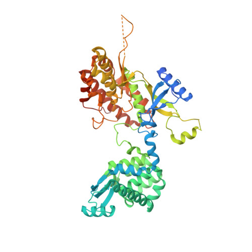
The crystal structure of the phosphatidylinositol 4-kinase II alpha.

Baumlova, A.Chalupska, D.Rozycki, B.Jovic, M.Wisniewski, E.Klima, M.Dubankova, A.Kloer, D.P.Nencka, R.Balla, T.Boura, E.

(2014) EMBO Rep 15: 1085-1092

  • DOI: https://doi.org/10.15252/embr.201438841
  • Primary Citation Related Structures: 
    4PLA

  • PubMed Abstract: 

    Phosphoinositides are a class of phospholipids generated by the action of phosphoinositide kinases with key regulatory functions in eukaryotic cells. Here, we present the atomic structure of phosphatidylinositol 4-kinase type IIα (PI4K IIα), in complex with ATP solved by X-ray crystallography at 2.8 Å resolution. The structure revealed a non-typical kinase fold that could be divided into N- and C-lobes with the ATP binding groove located in between. Surprisingly, a second ATP was found in a lateral hydrophobic pocket of the C-lobe. Molecular simulations and mutagenesis analysis revealed the membrane binding mode and the putative function of the hydrophobic pocket. Taken together, our results suggest a mechanism of PI4K IIα recruitment, regulation, and function at the membrane.


  • Organizational Affiliation
    • Institute of Organic Chemistry and Biochemistry AS CR, Prague, Czech Republic.

Macromolecule Content 

  • Total Structure Weight: 64.26 kDa 
  • Atom Count: 3,951 
  • Modeled Residue Count: 483 
  • Deposited Residue Count: 556 
  • Unique protein chains: 1

Macromolecules

Find similar proteins by:|  3D Structure
Entity ID: 1
MoleculeChains  Sequence LengthOrganismDetailsImage
Chimera protein of Phosphatidylinositol 4-kinase type 2-alpha and Lysozyme556Homo sapiensTequatrovirus T4Mutation(s): 2 
Gene Names: PI4K2AeT4Tp126
EC: 2.7.1.67 (PDB Primary Data), 3.2.1.17 (PDB Primary Data)
Membrane Entity: Yes 
UniProt & NIH Common Fund Data Resources
Find proteins for D9IEF7 (Enterobacteria phage T4)
Explore D9IEF7 
Go to UniProtKB:  D9IEF7
Find proteins for Q9BTU6 (Homo sapiens)
Explore Q9BTU6 
Go to UniProtKB:  Q9BTU6
PHAROS:  Q9BTU6
GTEx:  ENSG00000155252 
Entity Groups
Sequence Clusters30% Identity50% Identity70% Identity90% Identity95% Identity100% Identity
UniProt GroupsQ9BTU6D9IEF7
Sequence Annotations
Expand
Reference Sequence

Experimental Data & Validation

Experimental Data

  • Method: X-RAY DIFFRACTION
  • Resolution: 2.77 Å
  • R-Value Free:  0.251 (Depositor), 0.257 (DCC) 
  • R-Value Work:  0.211 (Depositor), 0.220 (DCC) 
  • R-Value Observed: 0.213 (Depositor) 
Space Group: P 21 21 2
Unit Cell:
Length ( Å )Angle ( ˚ )
a = 104.744α = 90
b = 79.539β = 90
c = 78.704γ = 90
Software Package:
Software NamePurpose
PHENIXrefinement

Structure Validation

View Full Validation Report


Ligand Structure Quality Assessment 


Entry History 

& Funding Information

Deposition Data


Funding OrganizationLocationGrant Number
MarieCurie FP7-PEOPLE-2012-CIG333916

Revision History  (Full details and data files)

  • Version 1.0: 2014-09-10
    Type: Initial release
  • Version 1.1: 2014-10-15
    Changes: Database references
  • Version 1.2: 2015-02-04
    Changes: Derived calculations
  • Version 1.3: 2023-12-27
    Changes: Data collection, Database references, Derived calculations, Other, Refinement description, Source and taxonomy