4PJ1

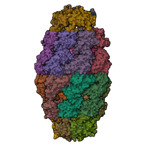
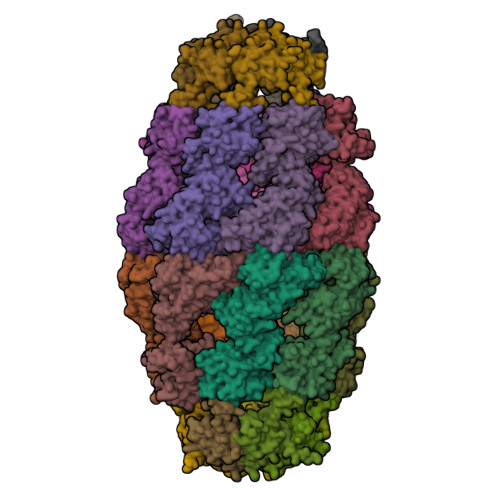
Crystal structure of the human mitochondrial chaperonin symmetrical 'football' complex


Experimental Data Snapshot

  • Method: X-RAY DIFFRACTION
  • Resolution: 3.15 Å
  • R-Value Free: 0.270 
  • R-Value Work: 0.241 
  • R-Value Observed: 0.242 

Starting Model: experimental
View more details

wwPDB Validation   3D Report Full Report


Ligand Structure Quality Assessment 


This is version 1.4 of the entry. See complete history


Literature

Crystal structure of the human mitochondrial chaperonin symmetrical football complex.

Nisemblat, S.Yaniv, O.Parnas, A.Frolow, F.Azem, A.

(2015) Proc Natl Acad Sci U S A 112: 6044-6049

  • DOI: https://doi.org/10.1073/pnas.1411718112
  • Primary Citation of Related Structures:  
    4PJ1

  • PubMed Abstract: 

    Human mitochondria harbor a single type I chaperonin system that is generally thought to function via a unique single-ring intermediate. To date, no crystal structure has been published for any mammalian type I chaperonin complex. In this study, we describe the crystal structure of a football-shaped, double-ring human mitochondrial chaperonin complex at 3.15 Å, which is a novel intermediate, likely representing the complex in an early stage of dissociation. Interestingly, the mitochondrial chaperonin was captured in a state that exhibits subunit asymmetry within the rings and nucleotide symmetry between the rings. Moreover, the chaperonin tetradecamers show a different interring subunit arrangement when compared to GroEL. Our findings suggest that the mitochondrial chaperonins use a mechanism that is distinct from the mechanism of the well-studied Escherichia coli system.


  • Organizational Affiliation

    Departments of Biochemistry and Molecular Biology and The Daniella Rich Institute for Structural Biology, The George S. Wise Faculty of Life Sciences, Tel Aviv University, Tel Aviv 69978, Israel.


Macromolecules
Find similar proteins by:  (by identity cutoff)  |  3D Structure
Entity ID: 1
MoleculeChains Sequence LengthOrganismDetailsImage
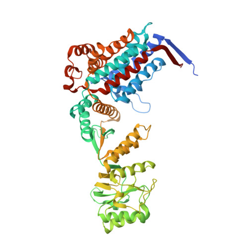
60 kDa heat shock protein, mitochondrial
A, B, C, D, E
A, B, C, D, E, F, G, H, I, J, K, L, M, N
558Homo sapiensMutation(s): 1 
Gene Names: HSPD1HSP60
EC: 5.6.1.7
UniProt & NIH Common Fund Data Resources
Find proteins for P10809 (Homo sapiens)
Explore P10809 
Go to UniProtKB:  P10809
PHAROS:  P10809
GTEx:  ENSG00000144381 
Entity Groups  
Sequence Clusters30% Identity50% Identity70% Identity90% Identity95% Identity100% Identity
UniProt GroupP10809
Sequence Annotations
Expand
  • Reference Sequence
Find similar proteins by:  (by identity cutoff)  |  3D Structure
Entity ID: 2
MoleculeChains Sequence LengthOrganismDetailsImage
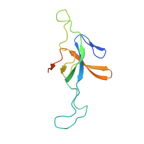
10 kDa heat shock protein, mitochondrial114Homo sapiensMutation(s): 0 
Gene Names: HSPE1
UniProt & NIH Common Fund Data Resources
Find proteins for P61604 (Homo sapiens)
Explore P61604 
Go to UniProtKB:  P61604
PHAROS:  P61604
GTEx:  ENSG00000115541 
Entity Groups  
Sequence Clusters30% Identity50% Identity70% Identity90% Identity95% Identity100% Identity
UniProt GroupP61604
Sequence Annotations
Expand
  • Reference Sequence
Small Molecules
Ligands 2 Unique
IDChains Name / Formula / InChI Key2D Diagram3D Interactions
ADP
Query on ADP

Download Ideal Coordinates CCD File 
AB [auth M]
CA [auth A]
CB [auth N]
EA [auth B]
GA [auth C]
AB [auth M],
CA [auth A],
CB [auth N],
EA [auth B],
GA [auth C],
IA [auth D],
KA [auth E],
MA [auth F],
OA [auth G],
QA [auth H],
SA [auth I],
UA [auth J],
WA [auth K],
YA [auth L]
ADENOSINE-5'-DIPHOSPHATE
C10 H15 N5 O10 P2
XTWYTFMLZFPYCI-KQYNXXCUSA-N
MG
Query on MG

Download Ideal Coordinates CCD File 
BB [auth M]
DA [auth A]
DB [auth N]
FA [auth B]
HA [auth C]
BB [auth M],
DA [auth A],
DB [auth N],
FA [auth B],
HA [auth C],
JA [auth D],
LA [auth E],
NA [auth F],
PA [auth G],
RA [auth H],
TA [auth I],
VA [auth J],
XA [auth K],
ZA [auth L]
MAGNESIUM ION
Mg
JLVVSXFLKOJNIY-UHFFFAOYSA-N
Experimental Data & Validation

Experimental Data

  • Method: X-RAY DIFFRACTION
  • Resolution: 3.15 Å
  • R-Value Free: 0.270 
  • R-Value Work: 0.241 
  • R-Value Observed: 0.242 
  • Space Group: P 41 21 2
Unit Cell:
Length ( Å )Angle ( ˚ )
a = 199.1α = 90
b = 199.1β = 90
c = 627.39γ = 90
Software Package:
Software NamePurpose
XDSdata reduction
PDB_EXTRACTdata extraction
MOLREPphasing
PHENIXrefinement
XSCALEdata scaling
Aimlessdata scaling

Structure Validation

View Full Validation Report


Ligand Structure Quality Assessment 


Entry History & Funding Information

Deposition Data


Funding OrganizationLocationGrant Number
Morasha ISFIsrael1902/08
Eshkol Fellowship to Shahar NissemblatIsrael--

Revision History  (Full details and data files)

  • Version 1.0: 2015-04-29
    Type: Initial release
  • Version 1.1: 2015-05-13
    Changes: Derived calculations
  • Version 1.2: 2015-05-20
    Changes: Database references
  • Version 1.3: 2017-11-22
    Changes: Database references, Derived calculations, Refinement description, Source and taxonomy
  • Version 1.4: 2023-09-27
    Changes: Data collection, Database references, Refinement description