4PIQ | pdb_00004piq

Crystal structure of human adenovirus 8 protease with a nitrile inhibitor


Experimental Data Snapshot

  • Method: X-RAY DIFFRACTION
  • Resolution: 2.07 Å
  • R-Value Free: 
    0.319 (Depositor), 0.339 (DCC) 
  • R-Value Work: 
    0.242 (Depositor), 0.251 (DCC) 
  • R-Value Observed: 
    0.246 (Depositor) 

wwPDB Validation 3D Report Full Report

Validation slider image for 4PIQ

Ligand Structure Quality Assessment 


This is version 1.3 of the entry. See complete history

Literature

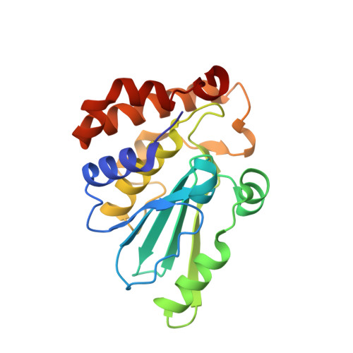
Discovery and structure-based optimization of adenain inhibitors.

Mac Sweeney, A.Grosche, P.Ellis, D.Combrink, K.Erbel, P.Hughes, N.Sirockin, F.Melkko, S.Bernardi, A.Ramage, P.Jarousse, N.Altmann, E.

(2014) ACS Med Chem Lett 5: 937-941

  • DOI: https://doi.org/10.1021/ml500224t
  • Primary Citation Related Structures: 
    4PID, 4PIE, 4PIQ, 4PIS

  • PubMed Abstract: 

    The cysteine protease adenain is the essential protease of adenovirus and, as such, represents a promising target for the treatment of ocular and other adenoviral infections. Through a concise two-pronged hit discovery approach we identified tetrapeptide nitrile 1 and pyrimidine nitrile 2 as complementary starting points for adenain inhibition. These hits enabled the first high-resolution X-ray cocrystal structures of adenain with inhibitors bound and revealed the binding mode of 1 and 2. The screening hits were optimized by a structure-guided medicinal chemistry strategy into low nanomolar drug-like inhibitors of adenain.


  • Organizational Affiliation
    • Novartis Institute for Biomedical Research , Novartis Campus, CH-4002 Basel, Switzerland.

Macromolecule Content 

  • Total Structure Weight: 24.77 kDa 
  • Atom Count: 1,841 
  • Modeled Residue Count: 215 
  • Deposited Residue Count: 215 
  • Unique protein chains: 2

Macromolecules

Find similar proteins by:|  3D Structure
Entity ID: 1
MoleculeChains  Sequence LengthOrganismDetailsImage
Protease204Human adenovirus D8Mutation(s): 0 
Gene Names: L3
EC: 3.4.22.39
UniProt
Find proteins for B9A5C1 (Human adenovirus D serotype 8)
Explore B9A5C1 
Go to UniProtKB:  B9A5C1
Entity Groups
Sequence Clusters30% Identity50% Identity70% Identity90% Identity95% Identity100% Identity
UniProt GroupB9A5C1
Sequence Annotations
Expand
Reference Sequence
Find similar proteins by:  Sequence   |   3D Structure  
Entity ID: 2
MoleculeChains  Sequence LengthOrganismDetailsImage
PVI11Human adenovirus D8Mutation(s): 0 
UniProt
Find proteins for B9A5B9 (Human adenovirus D serotype 8)
Explore B9A5B9 
Go to UniProtKB:  B9A5B9
Entity Groups
Sequence Clusters30% Identity50% Identity70% Identity90% Identity95% Identity100% Identity
UniProt GroupB9A5B9
Sequence Annotations
Expand
Reference Sequence

Small Molecules

Ligands 1 Unique
IDChains Name / Formula / InChI Key2D Diagram3D Interactions
3FS

Query on 3FS


Download:Ideal Coordinates CCD File
C [auth A]N-[(3,5-dichlorophenyl)acetyl]-L-threonyl-N-[(2Z)-2-iminoethyl]glycinamide
C16 H20 Cl2 N4 O4
MDMRSQQSLSAMBM-QMIWAGOPSA-N

Experimental Data & Validation

Experimental Data

  • Method: X-RAY DIFFRACTION
  • Resolution: 2.07 Å
  • R-Value Free:  0.319 (Depositor), 0.339 (DCC) 
  • R-Value Work:  0.242 (Depositor), 0.251 (DCC) 
  • R-Value Observed: 0.246 (Depositor) 
Space Group: P 32 2 1
Unit Cell:
Length ( Å )Angle ( ˚ )
a = 42.269α = 90
b = 42.269β = 90
c = 193.784γ = 120
Software Package:
Software NamePurpose
XDSdata reduction
PDB_EXTRACTdata extraction
XSCALEdata scaling
BUSTERrefinement

Structure Validation

View Full Validation Report


Ligand Structure Quality Assessment 


Entry History 

Revision History  (Full details and data files)

  • Version 1.0: 2014-09-10
    Type: Initial release
  • Version 1.1: 2017-08-23
    Changes: Data collection, Derived calculations, Other, Refinement description, Source and taxonomy
  • Version 1.2: 2023-12-27
    Changes: Data collection, Database references, Refinement description
  • Version 1.3: 2024-11-06
    Changes: Structure summary