4PHF

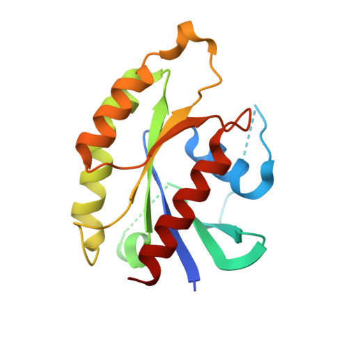
Crystal structure of Ypt7 covalently modified with GDP


Experimental Data Snapshot

  • Method: X-RAY DIFFRACTION
  • Resolution: 1.95 Å
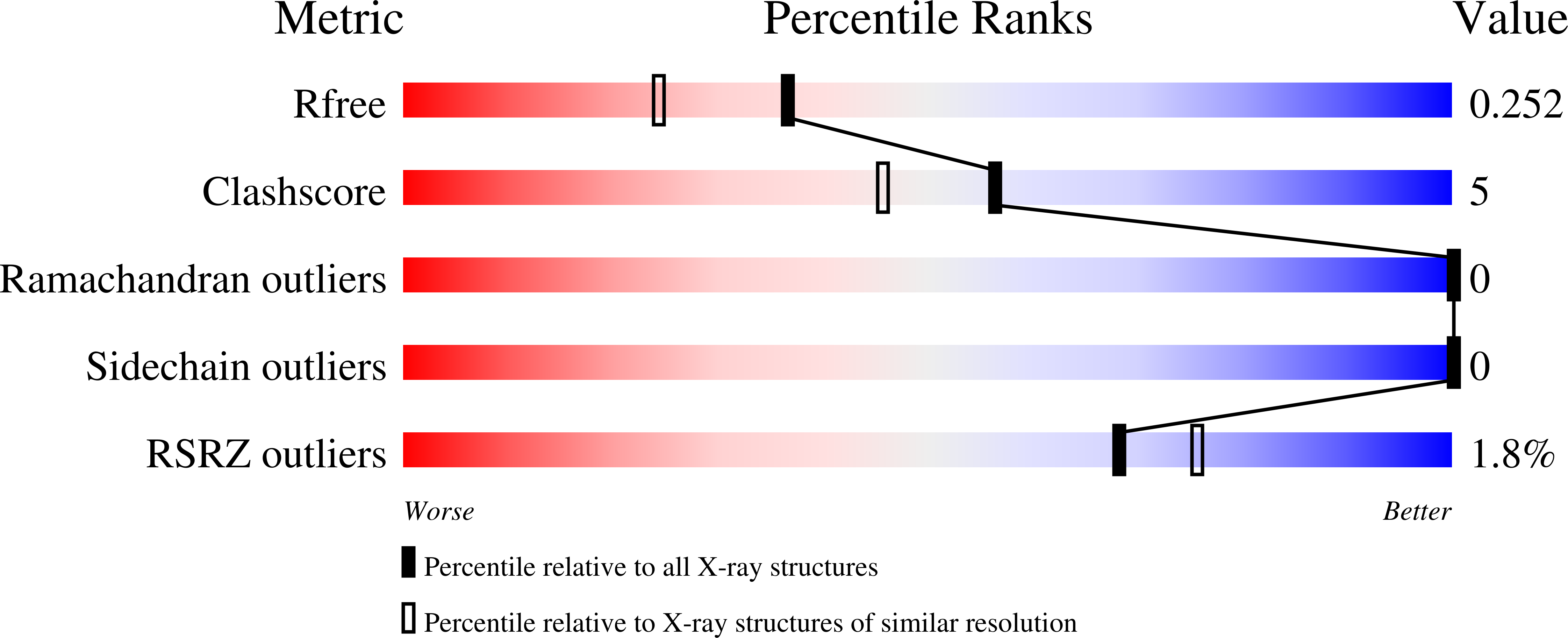
  • R-Value Free: 0.245 
  • R-Value Work: 0.196 
  • R-Value Observed: 0.199 

wwPDB Validation   3D Report Full Report


Ligand Structure Quality Assessment 

Created with Raphaël 2.3.0Worse 01 BetterLigand structure goodness of fit to experimental dataBest fitted 2UHClick on this verticalbar to view details

This is version 1.6 of the entry. See complete history


Literature

Locking GTPases covalently in their functional states.

Wiegandt, D.Vieweg, S.Hofmann, F.Koch, D.Li, F.Wu, Y.W.Itzen, A.Muller, M.P.Goody, R.S.

(2015) Nat Commun 6: 7773-7773

  • DOI: https://doi.org/10.1038/ncomms8773
  • Primary Citation of Related Structures:  
    4PHF, 4PHG, 4PHH

  • PubMed Abstract: 

    GTPases act as key regulators of many cellular processes by switching between active (GTP-bound) and inactive (GDP-bound) states. In many cases, understanding their mode of action has been aided by artificially stabilizing one of these states either by designing mutant proteins or by complexation with non-hydrolysable GTP analogues. Because of inherent disadvantages in these approaches, we have developed acryl-bearing GTP and GDP derivatives that can be covalently linked with strategically placed cysteines within the GTPase of interest. Binding studies with GTPase-interacting proteins and X-ray crystallography analysis demonstrate that the molecular properties of the covalent GTPase-acryl-nucleotide adducts are a faithful reflection of those of the corresponding native states and are advantageously permanently locked in a defined nucleotide (that is active or inactive) state. In a first application, in vivo experiments using covalently locked Rab5 variants provide new insights into the mechanism of correct intracellular localization of Rab proteins.


  • Organizational Affiliation

    Department of Physical Biochemistry, Max Planck Institute of Molecular Physiology, Otto-Hahn-Strasse 11, 44227 Dortmund, Germany.


Macromolecules
Find similar proteins by:  (by identity cutoff)  |  3D Structure
Entity ID: 1
MoleculeChains Sequence LengthOrganismDetailsImage
GTP-binding protein YPT7184Saccharomyces cerevisiae S288CMutation(s): 1 
Gene Names: YPT7VAM4YML001WYM8270.02
UniProt
Find proteins for P32939 (Saccharomyces cerevisiae (strain ATCC 204508 / S288c))
Explore P32939 
Go to UniProtKB:  P32939
Entity Groups  
Sequence Clusters30% Identity50% Identity70% Identity90% Identity95% Identity100% Identity
UniProt GroupP32939
Sequence Annotations
Expand
  • Reference Sequence
Experimental Data & Validation

Experimental Data

  • Method: X-RAY DIFFRACTION
  • Resolution: 1.95 Å
  • R-Value Free: 0.245 
  • R-Value Work: 0.196 
  • R-Value Observed: 0.199 
  • Space Group: P 21 21 21
Unit Cell:
Length ( Å )Angle ( ˚ )
a = 49.31α = 90
b = 54.9β = 90
c = 60.47γ = 90
Software Package:
Software NamePurpose
REFMACrefinement

Structure Validation

View Full Validation Report


Ligand Structure Quality Assessment 

Created with Raphaël 2.3.0Worse 01 BetterLigand structure goodness of fit to experimental dataBest fitted 2UHClick on this verticalbar to view details

Entry History & Funding Information

Deposition Data


Funding OrganizationLocationGrant Number
German Research Foundation (DFG)GermanySFB642, grant A4
German Research Foundation (DFG)GermanySFB1035, project B05

Revision History  (Full details and data files)

  • Version 1.0: 2014-05-28
    Type: Initial release
  • Version 1.1: 2015-02-04
    Changes: Derived calculations
  • Version 1.2: 2016-08-31
    Changes: Database references
  • Version 1.3: 2017-09-27
    Changes: Author supporting evidence, Derived calculations
  • Version 1.4: 2020-01-08
    Changes: Author supporting evidence
  • Version 1.5: 2023-12-27
    Changes: Data collection, Database references, Derived calculations
  • Version 1.6: 2024-10-23
    Changes: Structure summary