4PFW

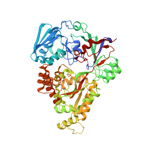
Crystal structure of mannohexaose bound oligopeptide ABC transporter, periplasmic oligopeptide-binding protein (TM1226) from THERMOTOGA MARITIMA at 2.2 A resolution


Experimental Data Snapshot

  • Method: X-RAY DIFFRACTION
  • Resolution: 2.20 Å
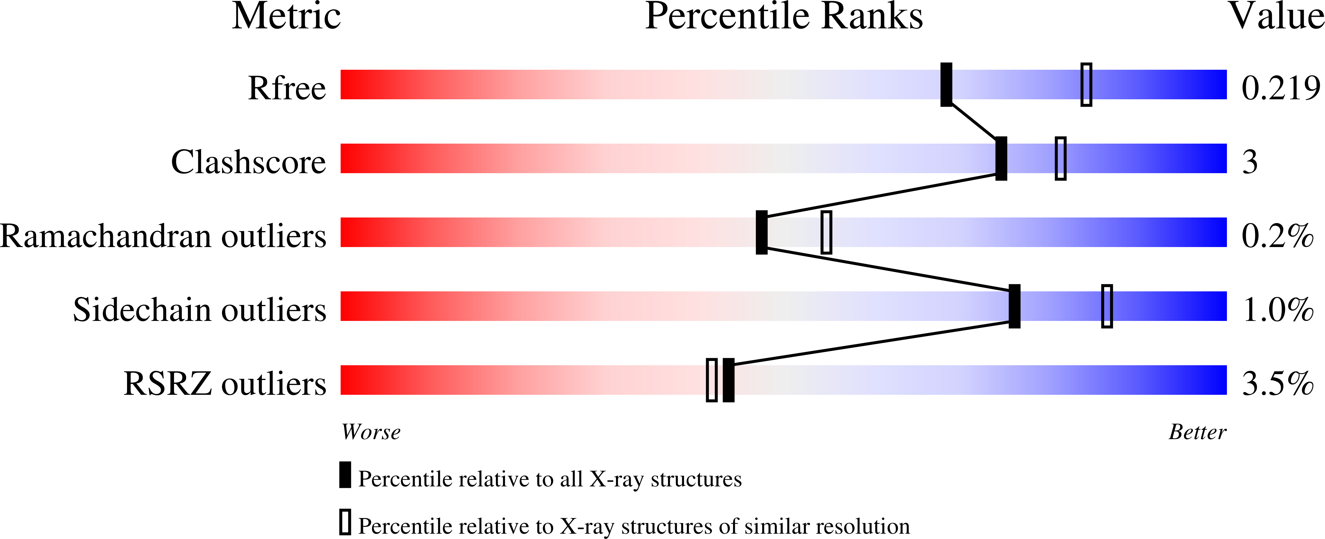
  • R-Value Free: 0.218 
  • R-Value Work: 0.164 
  • R-Value Observed: 0.167 

wwPDB Validation   3D Report Full Report


This is version 3.1 of the entry. See complete history


Literature

Duplication of Genes in an ATP-binding Cassette Transport System Increases Dynamic Range While Maintaining Ligand Specificity.

Ghimire-Rijal, S.Lu, X.Myles, D.A.Cuneo, M.J.

(2014) J Biol Chem 289: 30090-30100

  • DOI: https://doi.org/10.1074/jbc.M114.590992
  • Primary Citation of Related Structures:  
    4PFT, 4PFU, 4PFW, 4PFY

  • PubMed Abstract: 

    Many bacteria exist in a state of feast or famine where high nutrient availability leads to periods of growth followed by nutrient scarcity and growth stagnation. To adapt to the constantly changing nutrient flux, metabolite acquisition systems must be able to function over a broad range. This, however, creates difficulties as nutrient concentrations vary over many orders of magnitude, requiring metabolite acquisition systems to simultaneously balance ligand specificity and the dynamic range in which a response to a metabolite is elicited. Here we present how a gene duplication of a periplasmic binding protein in a mannose ATP-binding cassette transport system potentially resolves this dilemma through gene functionalization. Determination of ligand binding affinities and specificities of the gene duplicates with fluorescence and circular dichroism demonstrates that although the binding specificity is maintained the Kd values for the same ligand differ over three orders of magnitude. These results suggest that this metabolite acquisition system can transport ligand at both low and high environmental concentrations while preventing saturation with related and less preferentially metabolized compounds. The x-ray crystal structures of the β-mannose-bound proteins help clarify the structural basis of gene functionalization and reveal that affinity and specificity are potentially encoded in different regions of the binding site. These studies suggest a possible functional role and adaptive advantage for the presence of two periplasmic-binding proteins in ATP-binding cassette transport systems and a way bacteria can adapt to varying nutrient flux through functionalization of gene duplicates.


  • Organizational Affiliation

    From the Neutron Sciences Directorate, Oak Ridge National Laboratory, Oak Ridge, Tennessee 37831.


Macromolecules
Find similar proteins by:  (by identity cutoff)  |  3D Structure
Entity ID: 1
MoleculeChains Sequence LengthOrganismDetailsImage
ABC transporter substrate-binding protein
A, B
547Thermotoga maritima MSB8Mutation(s): 0 
Gene Names: THEMA_08200Tmari_1233
Entity Groups  
Sequence Clusters30% Identity50% Identity70% Identity90% Identity95% Identity100% Identity
Sequence Annotations
Expand
  • Reference Sequence
Oligosaccharides

Help

Entity ID: 2
MoleculeChains Length2D Diagram Glycosylation3D Interactions
beta-D-mannopyranose-(1-4)-beta-D-mannopyranose-(1-4)-beta-D-mannopyranose-(1-4)-beta-D-mannopyranose-(1-4)-alpha-D-mannopyranose-(1-4)-alpha-D-mannopyranose
C, D
6N/A
Glycosylation Resources
GlyTouCan:  G32802UG
GlyCosmos:  G32802UG
GlyGen:  G32802UG
Small Molecules
Ligands 3 Unique
IDChains Name / Formula / InChI Key2D Diagram3D Interactions
SO4
Query on SO4

Download Ideal Coordinates CCD File 
F [auth A],
G [auth A],
H [auth A],
I [auth A],
N [auth B]
SULFATE ION
O4 S
QAOWNCQODCNURD-UHFFFAOYSA-L
GOL
Query on GOL

Download Ideal Coordinates CCD File 
K [auth B],
L [auth B],
M [auth B]
GLYCEROL
C3 H8 O3
PEDCQBHIVMGVHV-UHFFFAOYSA-N
MG
Query on MG

Download Ideal Coordinates CCD File 
E [auth A],
J [auth B]
MAGNESIUM ION
Mg
JLVVSXFLKOJNIY-UHFFFAOYSA-N
Experimental Data & Validation

Experimental Data

  • Method: X-RAY DIFFRACTION
  • Resolution: 2.20 Å
  • R-Value Free: 0.218 
  • R-Value Work: 0.164 
  • R-Value Observed: 0.167 
  • Space Group: C 2 2 21
Unit Cell:
Length ( Å )Angle ( ˚ )
a = 99.548α = 90
b = 194.227β = 90
c = 159.425γ = 90
Software Package:
Software NamePurpose
PHENIXrefinement

Structure Validation

View Full Validation Report


Entry History & Funding Information

Deposition Data


Funding OrganizationLocationGrant Number
Department of Energy (DOE, United States)United States--

Revision History  (Full details and data files)

  • Version 1.0: 2014-09-17
    Type: Initial release
  • Version 1.1: 2014-09-24
    Changes: Database references
  • Version 1.2: 2014-11-05
    Changes: Database references
  • Version 2.0: 2017-09-27
    Changes: Advisory, Atomic model, Author supporting evidence, Database references, Derived calculations, Other, Source and taxonomy, Structure summary
  • Version 2.1: 2019-12-04
    Changes: Author supporting evidence
  • Version 3.0: 2020-07-29
    Type: Remediation
    Reason: Carbohydrate remediation
    Changes: Atomic model, Data collection, Derived calculations, Refinement description, Structure summary
  • Version 3.1: 2023-12-27
    Changes: Data collection, Database references, Derived calculations, Structure summary