4PF5 | pdb_00004pf5

Crystal structure of Concanavalin A complexed with a synthetic derivative of high-mannose chain


Experimental Data Snapshot

  • Method: X-RAY DIFFRACTION
  • Resolution: 2.04 Å
  • R-Value Free: 
    0.227 (Depositor), 0.235 (DCC) 
  • R-Value Work: 
    0.178 (Depositor), 0.191 (DCC) 
  • R-Value Observed: 
    0.181 (Depositor) 

wwPDB Validation 3D Report Full Report

Validation slider image for 4PF5

Ligand Structure Quality Assessment 


This is version 1.3 of the entry. See complete history

Literature

Synthesis of High-Mannose Oligosaccharide Analogues through Click Chemistry: True Functional Mimics of Their Natural Counterparts Against Lectins?

Francois-Heude, M.Mendez-Ardoy, A.Cendret, V.Lafite, P.Daniellou, R.Ortiz Mellet, C.Garcia Fernandez, J.M.Moreau, V.Djedaini-Pilard, F.

(2015) Chemistry 21: 1978-1991

  • DOI: https://doi.org/10.1002/chem.201405481
  • Primary Citation Related Structures: 
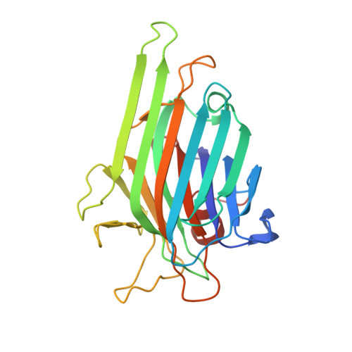
    4PF5

  • PubMed Abstract: 

    Terminal "high-mannose oligosaccharides" are involved in a broad range of biological and pathological processes, from sperm-egg fusion to influenza and human immunodeficiency virus infections. In spite of many efforts, their synthesis continues to be very challenging and actually represents a major bottleneck in the field. Whereas multivalent presentation of mannopyranosyl motifs onto a variety of scaffolds has proven to be a successful way to interfere in recognition processes involving high-mannose oligosaccharides, such constructs fail at reproducing the subtle differences in affinity towards the variety of protein receptors (lectins) and antibodies susceptible to binding to the natural ligands. Here we report a family of functional high-mannose oligosaccharide mimics that reproduce not only the terminal mannopyranosyl display, but also the core structure and the branching pattern, by replacing some inner mannopyranosyl units with triazole rings. Such molecular design can be implemented by exploiting "click" ligation strategies, resulting in a substantial reduction of synthetic cost. The binding affinities of the new "click" high-mannose oligosaccharide mimics towards two mannose specific lectins, namely the plant lectin concanavalin A (ConA) and the human macrophage mannose receptor (rhMMR), have been studied by enzyme-linked lectin assays and found to follow identical trends to those observed for the natural oligosaccharide counterparts. Calorimetric determinations against ConA, and X-ray structural data support the conclusion that these compounds are not just another family of multivalent mannosides, but real "structural mimics" of the high-mannose oligosaccharides.


  • Organizational Affiliation
    • LG-2A FRE-CNRS 3517, Institut de Chimie de Picardie, Université de Picardie Jules Verne, 33 Rue Saint-Leu, 80039 Amiens Cedex 1 (France).

Macromolecule Content 

  • Total Structure Weight: 52.11 kDa 
  • Atom Count: 3,902 
  • Modeled Residue Count: 474 
  • Deposited Residue Count: 474 
  • Unique protein chains: 1

Macromolecules

Find similar proteins by:|  3D Structure
Entity ID: 1
MoleculeChains  Sequence LengthOrganismDetailsImage
Concanavalin-A
A, B
237Canavalia ensiformisMutation(s): 0 
UniProt
Find proteins for P02866 (Canavalia ensiformis)
Explore P02866 
Go to UniProtKB:  P02866
Entity Groups
Sequence Clusters30% Identity50% Identity70% Identity90% Identity95% Identity100% Identity
UniProt GroupP02866
Sequence Annotations
Expand
Reference Sequence

Small Molecules

Ligands 3 Unique
IDChains Name / Formula / InChI Key2D Diagram3D Interactions
M3N

Query on M3N


Download:Ideal Coordinates CCD File
E [auth A],
G [auth B]
4-(hydroxymethyl)-1-(alpha-D-mannopyranosyl)-1H-1,2,3-triazole
C9 H15 N3 O6
BHGZOVXPACYZLI-DFTQBPQZSA-N
EDO

Query on EDO


Download:Ideal Coordinates CCD File
F [auth A],
J [auth B]
1,2-ETHANEDIOL
C2 H6 O2
LYCAIKOWRPUZTN-UHFFFAOYSA-N
MN

Query on MN


Download:Ideal Coordinates CCD File
C [auth A],
D [auth A],
H [auth B],
I [auth B]
MANGANESE (II) ION
Mn
WAEMQWOKJMHJLA-UHFFFAOYSA-N

Experimental Data & Validation

Experimental Data

  • Method: X-RAY DIFFRACTION
  • Resolution: 2.04 Å
  • R-Value Free:  0.227 (Depositor), 0.235 (DCC) 
  • R-Value Work:  0.178 (Depositor), 0.191 (DCC) 
  • R-Value Observed: 0.181 (Depositor) 
Space Group: H 3 2
Unit Cell:
Length ( Å )Angle ( ˚ )
a = 125.901α = 90
b = 125.901β = 90
c = 181.972γ = 120
Software Package:
Software NamePurpose
XDSdata reduction
SCALAdata scaling
PHASERphasing
PHENIXrefinement
PDB_EXTRACTdata extraction

Structure Validation

View Full Validation Report


Ligand Structure Quality Assessment 


Entry History 

Deposition Data

Revision History  (Full details and data files)

  • Version 1.0: 2014-12-17
    Type: Initial release
  • Version 1.1: 2015-01-28
    Changes: Database references
  • Version 1.2: 2020-07-29
    Type: Remediation
    Reason: Carbohydrate remediation
    Changes: Data collection, Derived calculations
  • Version 1.3: 2024-05-08
    Changes: Data collection, Database references, Structure summary