4PD3 | pdb_00004pd3

Crystal Structure of Rigor-Like Human Nonmuscle Myosin-2B


Experimental Data Snapshot

  • Method: X-RAY DIFFRACTION
  • Resolution: 2.84 Å
  • R-Value Free: 
    0.288 (Depositor), 0.286 (DCC) 
  • R-Value Work: 
    0.253 (Depositor), 0.256 (DCC) 
  • R-Value Observed: 
    0.254 (Depositor) 

wwPDB Validation 3D Report Full Report

Validation slider image for 4PD3

This is version 2.1 of the entry. See complete history

Literature

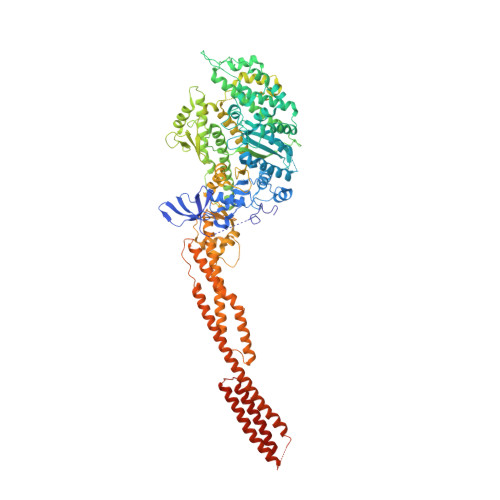
Crystal structure of the rigor-like human non-muscle myosin-2 motor domain.

Munnich, S.Pathan-Chhatbar, S.Manstein, D.J.

(2014) FEBS Lett 588: 4754-4760

  • DOI: https://doi.org/10.1016/j.febslet.2014.11.007
  • Primary Citation Related Structures: 
    4PD3

  • PubMed Abstract: 

    We determined the crystal structure of the motor domain of human non-muscle myosin 2B (NM-2B) in a nucleotide-free state and at a resolution of 2.8 Å. The structure shows the motor domain with an open active site and the large cleft that divides the 50 kDa domain in a closed state. Compared to other rigor-like myosin motor domain structures, our structure shows subtle but significant conformational changes in regions important for actin binding and mechanochemical coupling. Moreover, our crystal structure helps to rationalize the impact of myosin, heavy chain 9 (MYH9)-related disease mutations Arg709Cys and Arg709His on the kinetic and functional properties of NM-2B and of the closely related non-muscle myosin 2A (NM-2A).


  • Organizational Affiliation
    • Institute for Biophysical Chemistry, Hannover Medical School, Hannover, Germany.

Macromolecule Content 

  • Total Structure Weight: 237.43 kDa 
  • Atom Count: 14,828 
  • Modeled Residue Count: 1,852 
  • Deposited Residue Count: 2,064 
  • Unique protein chains: 1

Macromolecules

Find similar proteins by:|  3D Structure
Entity ID: 1
MoleculeChains  Sequence LengthOrganismDetailsImage
Nonmuscle myosin heavy chain B, Alpha-actinin A Chimera Protein
A, B
1,032Homo sapiensDictyostelium discoideum
This entity is chimeric
Mutation(s): 0 
Gene Names: MYH10abpAactnADDB_G0268632
UniProt & NIH Common Fund Data Resources
Find proteins for P05095 (Dictyostelium discoideum)
Explore P05095 
Go to UniProtKB:  P05095
Find proteins for P35580 (Homo sapiens)
Explore P35580 
Go to UniProtKB:  P35580
PHAROS:  P35580
GTEx:  ENSG00000133026 
Entity Groups
Sequence Clusters30% Identity50% Identity70% Identity90% Identity95% Identity100% Identity
UniProt GroupsP35580P05095
Sequence Annotations
Expand
Reference Sequence

Experimental Data & Validation

Experimental Data

  • Method: X-RAY DIFFRACTION
  • Resolution: 2.84 Å
  • R-Value Free:  0.288 (Depositor), 0.286 (DCC) 
  • R-Value Work:  0.253 (Depositor), 0.256 (DCC) 
  • R-Value Observed: 0.254 (Depositor) 
Space Group: P 1 21 1
Unit Cell:
Length ( Å )Angle ( ˚ )
a = 65.85α = 90
b = 157.87β = 94.21
c = 143.48γ = 90
Software Package:
Software NamePurpose
PHENIXrefinement

Structure Validation

View Full Validation Report


Entry History 

Deposition Data

Revision History  (Full details and data files)

  • Version 1.0: 2014-11-12
    Type: Initial release
  • Version 1.1: 2014-12-17
    Changes: Database references
  • Version 1.2: 2014-12-24
    Changes: Database references
  • Version 1.3: 2015-02-04
    Changes: Derived calculations
  • Version 1.4: 2018-03-07
    Changes: Data collection, Database references, Derived calculations, Other, Source and taxonomy, Structure summary
  • Version 2.0: 2020-08-19
    Changes: Atomic model, Data collection, Refinement description, Source and taxonomy, Structure summary
  • Version 2.1: 2023-12-27
    Changes: Data collection, Database references