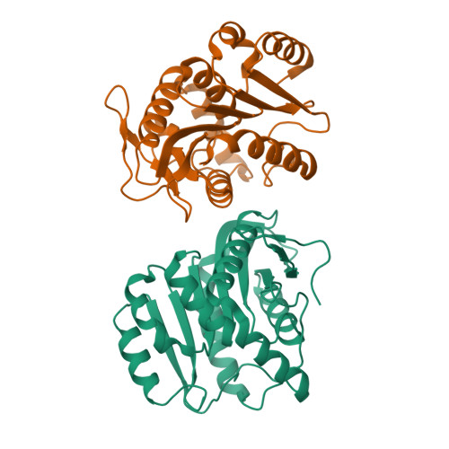
4P93

Structure of Dienelactone Hydrolase at 1.85 A resolution crystallised in the C2 space group


Experimental Data Snapshot

  • Method: X-RAY DIFFRACTION
  • Resolution: 1.85 Å
  • R-Value Free: 0.205 
  • R-Value Work: 0.173 
  • R-Value Observed: 0.175 

Starting Model: experimental
View more details

wwPDB Validation   3D Report Full Report


This is version 1.2 of the entry. See complete history


Literature

Crystallization of dienelactone hydrolase in two space groups: structural changes caused by crystal packing.

Porter, J.L.Carr, P.D.Collyer, C.A.Ollis, D.L.

(2014) Acta Crystallogr F Struct Biol Commun 70: 884-889

  • DOI: https://doi.org/10.1107/S2053230X1401108X
  • Primary Citation of Related Structures:  
    4P92, 4P93

  • PubMed Abstract: 

    Dienelactone hydrolase (DLH) is a monomeric protein with a simple α/β-hydrolase fold structure. It readily crystallizes in space group P2₁2₁2₁ from either a phosphate or ammonium sulfate precipitation buffer. Here, the structure of DLH at 1.85 Å resolution crystallized in space group C2 with two molecules in the asymmetric unit is reported. When crystallized in space group P2₁2₁2₁ DLH has either phosphates or sulfates bound to the protein in crucial locations, one of which is located in the active site, preventing substrate/inhibitor binding. Another is located on the surface of the enzyme coordinated by side chains from two different molecules. Crystallization in space group C2 from a sodium citrate buffer results in new crystallographic protein-protein interfaces. The protein backbone is highly similar, but new crystal contacts cause changes in side-chain orientations and in loop positioning. In regions not involved in crystal contacts, there is little change in backbone or side-chain configuration. The flexibility of surface loops and the adaptability of side chains are important factors enabling DLH to adapt and form different crystal lattices.


  • Organizational Affiliation

    Research School of Chemistry, Australian National University, Building 137, Sullivans Creek Road, Canberra, ACT 0200, Australia.


Macromolecules
Find similar proteins by:  (by identity cutoff)  |  3D Structure
Entity ID: 1
MoleculeChains Sequence LengthOrganismDetailsImage
Carboxymethylenebutenolidase
A, B
236Pseudomonas knackmussiiMutation(s): 1 
Gene Names: clcD
EC: 3.1.1.45
UniProt
Find proteins for P0A115 (Pseudomonas knackmussii (strain DSM 6978 / CCUG 54928 / LMG 23759 / B13))
Explore P0A115 
Go to UniProtKB:  P0A115
Entity Groups  
Sequence Clusters30% Identity50% Identity70% Identity90% Identity95% Identity100% Identity
UniProt GroupP0A115
Sequence Annotations
Expand
  • Reference Sequence
Experimental Data & Validation

Experimental Data

  • Method: X-RAY DIFFRACTION
  • Resolution: 1.85 Å
  • R-Value Free: 0.205 
  • R-Value Work: 0.173 
  • R-Value Observed: 0.175 
  • Space Group: C 1 2 1
Unit Cell:
Length ( Å )Angle ( ˚ )
a = 100.23α = 90
b = 49.05β = 94.3
c = 89.61γ = 90
Software Package:
Software NamePurpose
REFMACrefinement
XDSdata reduction
XSCALEdata scaling
PHASERphasing

Structure Validation

View Full Validation Report


Entry History 

Deposition Data

Revision History  (Full details and data files)

  • Version 1.0: 2014-07-09
    Type: Initial release
  • Version 1.1: 2014-10-15
    Changes: Database references
  • Version 1.2: 2023-12-20
    Changes: Data collection, Database references, Refinement description