4P8C | pdb_00004p8c

Crystal structure of M. tuberculosis DprE1 in complex with the non-covalent inhibitor QN127


Experimental Data Snapshot

  • Method: X-RAY DIFFRACTION
  • Resolution: 1.95 Å
  • R-Value Free: 
    0.239 (Depositor), 0.237 (DCC) 
  • R-Value Work: 
    0.202 (Depositor), 0.201 (DCC) 
  • R-Value Observed: 
    0.204 (Depositor) 

Starting Model: experimental
View more details

wwPDB Validation 3D Report Full Report

Validation slider image for 4P8C

Ligand Structure Quality Assessment 


This is version 1.4 of the entry. See complete history

Literature

2-Carboxyquinoxalines Kill Mycobacterium tuberculosis through Noncovalent Inhibition of DprE1.

Neres, J.Hartkoorn, R.C.Chiarelli, L.R.Gadupudi, R.Pasca, M.R.Mori, G.Venturelli, A.Savina, S.Makarov, V.Kolly, G.S.Molteni, E.Binda, C.Dhar, N.Ferrari, S.Brodin, P.Delorme, V.Landry, V.de Jesus Lopes Ribeiro, A.L.Farina, D.Saxena, P.Pojer, F.Carta, A.Luciani, R.Porta, A.Zanoni, G.De Rossi, E.Costi, M.P.Riccardi, G.Cole, S.T.

(2015) ACS Chem Biol 10: 705-714

  • DOI: https://doi.org/10.1021/cb5007163
  • Primary Citation Related Structures: 
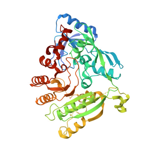
    4CVY, 4P8C, 4P8K, 4P8L, 4P8M, 4P8N, 4P8P, 4P8T, 4P8Y

  • PubMed Abstract: 

    Phenotypic screening of a quinoxaline library against replicating Mycobacterium tuberculosis led to the identification of lead compound Ty38c (3-((4-methoxybenzyl)amino)-6-(trifluoromethyl)quinoxaline-2-carboxylic acid). With an MIC99 and MBC of 3.1 μM, Ty38c is bactericidal and active against intracellular bacteria. To investigate its mechanism of action, we isolated mutants resistant to Ty38c and sequenced their genomes. Mutations were found in rv3405c, coding for the transcriptional repressor of the divergently expressed rv3406 gene. Biochemical studies clearly showed that Rv3406 decarboxylates Ty38c into its inactive keto metabolite. The actual target was then identified by isolating Ty38c-resistant mutants of an M. tuberculosis strain lacking rv3406. Here, mutations were found in dprE1, encoding the decaprenylphosphoryl-d-ribose oxidase DprE1, essential for biogenesis of the mycobacterial cell wall. Genetics, biochemical validation, and X-ray crystallography revealed Ty38c to be a noncovalent, noncompetitive DprE1 inhibitor. Structure-activity relationship studies generated a family of DprE1 inhibitors with a range of IC50's and bactericidal activity. Co-crystal structures of DprE1 in complex with eight different quinoxaline analogs provided a high-resolution interaction map of the active site of this extremely vulnerable target in M. tuberculosis.


  • Organizational Affiliation
    • †More Medicines for Tuberculosis (MM4TB) Consortium (www.mm4tb.org).

Macromolecule Content 

  • Total Structure Weight: 107.18 kDa 
  • Atom Count: 7,087 
  • Modeled Residue Count: 865 
  • Deposited Residue Count: 960 
  • Unique protein chains: 1

Macromolecules

Find similar proteins by:|  3D Structure
Entity ID: 1
MoleculeChains  Sequence LengthOrganismDetailsImage
Probable decaprenylphosphoryl-beta-D-ribose oxidase
A, B
480Mycobacterium tuberculosis H37RvMutation(s): 0 
Gene Names: dprE1Rv3790MT3898
EC: 1 (PDB Primary Data), 1.1.98.3 (UniProt)
UniProt
Find proteins for P9WJF1 (Mycobacterium tuberculosis (strain ATCC 25618 / H37Rv))
Explore P9WJF1 
Go to UniProtKB:  P9WJF1
Entity Groups
Sequence Clusters30% Identity50% Identity70% Identity90% Identity95% Identity100% Identity
UniProt GroupP9WJF1
Sequence Annotations
Expand
Reference Sequence

Small Molecules

Ligands 4 Unique
IDChains Name / Formula / InChI Key2D Diagram3D Interactions
FAD

Query on FAD


Download:Ideal Coordinates CCD File
C [auth A],
G [auth B]
FLAVIN-ADENINE DINUCLEOTIDE
C27 H33 N9 O15 P2
VWWQXMAJTJZDQX-UYBVJOGSSA-N
Y22

Query on Y22


Download:Ideal Coordinates CCD File
D [auth A],
H [auth B]
6-(trifluoromethyl)-3-{[4-(trifluoromethyl)benzyl]amino}quinoxaline-2-carboxylic acid
C18 H11 F6 N3 O2
FEJSMMIZTOMLEL-UHFFFAOYSA-N
2J3

Query on 2J3


Download:Ideal Coordinates CCD File
F [auth A](2R)-2-{[(2R)-2-{[(2R)-2-hydroxypropyl]oxy}propyl]oxy}propan-1-ol
C9 H20 O4
LCZVSXRMYJUNFX-IWSPIJDZSA-N
IMD

Query on IMD


Download:Ideal Coordinates CCD File
E [auth A]IMIDAZOLE
C3 H5 N2
RAXXELZNTBOGNW-UHFFFAOYSA-O
Binding Affinity Annotations 
IDSourceBinding Affinity
Y22 BindingDB:  4P8C IC50: 120 (nM) from 1 assay(s)

Experimental Data & Validation

Experimental Data

  • Method: X-RAY DIFFRACTION
  • Resolution: 1.95 Å
  • R-Value Free:  0.239 (Depositor), 0.237 (DCC) 
  • R-Value Work:  0.202 (Depositor), 0.201 (DCC) 
  • R-Value Observed: 0.204 (Depositor) 
Space Group: P 1 21 1
Unit Cell:
Length ( Å )Angle ( ˚ )
a = 77.678α = 90
b = 84.158β = 103.17
c = 80.905γ = 90
Software Package:
Software NamePurpose
XDSdata scaling
PHASERphasing
REFMACrefinement

Structure Validation

View Full Validation Report


Ligand Structure Quality Assessment 


Entry History 

Deposition Data

Revision History  (Full details and data files)

  • Version 1.0: 2014-12-10
    Type: Initial release
  • Version 1.1: 2015-02-04
    Changes: Derived calculations
  • Version 1.2: 2015-04-01
    Changes: Database references
  • Version 1.3: 2017-11-22
    Changes: Derived calculations, Other, Refinement description, Source and taxonomy
  • Version 1.4: 2023-09-27
    Changes: Data collection, Database references, Refinement description