4P7S | pdb_00004p7s

Crystal structure of PfMIF in complex with 4-(3-methoxy-5-methylphenoxy)-2-(4-methoxyphenyl)-6-methylpyridine


Experimental Data Snapshot

  • Method: X-RAY DIFFRACTION
  • Resolution: 2.87 Å
  • R-Value Free: 
    0.345 (Depositor), 0.354 (DCC) 
  • R-Value Work: 
    0.287 (Depositor), 0.292 (DCC) 
  • R-Value Observed: 
    0.290 (Depositor) 

wwPDB Validation 3D Report Full Report

Validation slider image for 4P7S

Ligand Structure Quality Assessment 


This is version 1.4 of the entry. See complete history

Literature

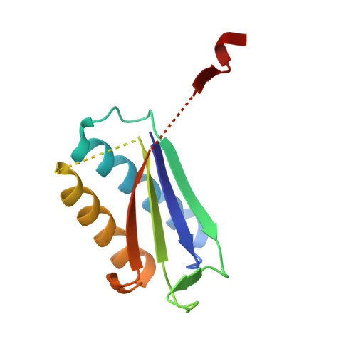
Crystallographic and Receptor Binding Characterization of Plasmodium falciparum Macrophage Migration Inhibitory Factor Complexed to Two Potent Inhibitors.

Pantouris, G.Rajasekaran, D.Garcia, A.B.Ruiz, V.G.Leng, L.Jorgensen, W.L.Bucala, R.Lolis, E.J.

(2014) J Med Chem 57: 8652-8656

  • DOI: https://doi.org/10.1021/jm501168q
  • Primary Citation Related Structures: 
    4P7M, 4P7S

  • PubMed Abstract: 

    We report the crystal structures of two inhibitors of Plasmodium falciparum macrophage migration inhibitory factor (PfMIF) with nanomolar Ki's, analyze their interactions with the active site of PfMIF, and provide explanations regarding their selectivity of PfMIF versus human MIF. These inhibitors were also found to selectively inhibit interactions between PfMIF and the human MIF receptor CD74. The results of this study provide the framework for the development of new therapeutics that target PfMIF.


  • Organizational Affiliation
    • Departments of Pharmacology, ‡Internal Medicine, §Chemistry, and the ∥Yale Cancer Center, Yale University , New Haven, Connecticut 06520-8066, United States.

Macromolecule Content 

  • Total Structure Weight: 38.67 kDa 
  • Atom Count: 2,136 
  • Modeled Residue Count: 297 
  • Deposited Residue Count: 342 
  • Unique protein chains: 1

Macromolecules

Find similar proteins by:|  3D Structure
Entity ID: 1
MoleculeChains  Sequence LengthOrganismDetailsImage
Macrophage migration inhibitory factor-like protein
A, B, C
114Plasmodium falciparumMutation(s): 0 
EC: 5.3.3.12 (UniProt), 5.3.2.1 (UniProt)
UniProt
Find proteins for Q6Q3H7 (Plasmodium falciparum)
Explore Q6Q3H7 
Go to UniProtKB:  Q6Q3H7
Entity Groups
Sequence Clusters30% Identity50% Identity70% Identity90% Identity95% Identity100% Identity
UniProt GroupQ6Q3H7
Sequence Annotations
Expand
Reference Sequence

Small Molecules

Ligands 1 Unique
IDChains Name / Formula / InChI Key2D Diagram3D Interactions
2OK

Query on 2OK


Download:Ideal Coordinates CCD File
D [auth B],
E [auth C]
4-(3-methoxy-5-methylphenoxy)-2-(4-methoxyphenyl)-6-methylpyridine
C21 H21 N O3
WAAJEIGWGSZNPQ-UHFFFAOYSA-N

Experimental Data & Validation

Experimental Data

  • Method: X-RAY DIFFRACTION
  • Resolution: 2.87 Å
  • R-Value Free:  0.345 (Depositor), 0.354 (DCC) 
  • R-Value Work:  0.287 (Depositor), 0.292 (DCC) 
  • R-Value Observed: 0.290 (Depositor) 
Space Group: I 2 2 2
Unit Cell:
Length ( Å )Angle ( ˚ )
a = 79.554α = 90
b = 80.138β = 90
c = 97.663γ = 90
Software Package:
Software NamePurpose
HKL-2000data collection
HKL-2000data reduction
HKL-2000data scaling
REFMACrefinement
PDB_EXTRACTdata extraction
PHASERphasing
DENZOdata reduction
SCALEPACKdata scaling

Structure Validation

View Full Validation Report


Ligand Structure Quality Assessment 


Entry History 

Deposition Data

Revision History  (Full details and data files)

  • Version 1.0: 2014-10-15
    Type: Initial release
  • Version 1.1: 2014-11-05
    Changes: Database references
  • Version 1.2: 2015-10-14
    Changes: Data collection
  • Version 1.3: 2017-11-22
    Changes: Derived calculations, Refinement description
  • Version 1.4: 2023-12-27
    Changes: Data collection, Database references