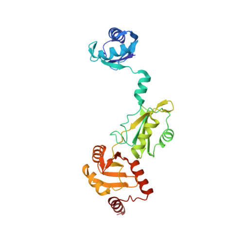
4P6Q | pdb_00004p6q

The crystal structure of the Split End protein SHARP adds a new layer of complexity to proteins containing RNA Recognition Motifs


Experimental Data Snapshot

  • Method: X-RAY DIFFRACTION
  • Resolution: 2.00 Å
  • R-Value Free: 
    0.238 (Depositor), 0.236 (DCC) 
  • R-Value Work: 
    0.192 (Depositor), 0.198 (DCC) 
  • R-Value Observed: 
    0.196 (Depositor) 

Starting Models: experimental
View more details

wwPDB Validation 3D Report Full Report

Validation slider image for 4P6Q

This is version 2.1 of the entry. See complete history

Literature

The crystal structure of the Split End protein SHARP adds a new layer of complexity to proteins containing RNA recognition motifs.

Arieti, F.Gabus, C.Tambalo, M.Huet, T.Round, A.Thore, S.

(2014) Nucleic Acids Res 42: 6742

  • DOI: https://doi.org/10.1093/nar/gku277
  • Primary Citation Related Structures: 
    4P6Q

  • PubMed Abstract: 

    The Split Ends (SPEN) protein was originally discovered in Drosophila in the late 1990s. Since then, homologous proteins have been identified in eukaryotic species ranging from plants to humans. Every family member contains three predicted RNA recognition motifs (RRMs) in the N-terminal region of the protein. We have determined the crystal structure of the region of the human SPEN homolog that contains these RRMs-the SMRT/HDAC1 Associated Repressor Protein (SHARP), at 2.0 Å resolution. SHARP is a co-regulator of the nuclear receptors. We demonstrate that two of the three RRMs, namely RRM3 and RRM4, interact via a highly conserved interface. Furthermore, we show that the RRM3-RRM4 block is the main platform mediating the stable association with the H12-H13 substructure found in the steroid receptor RNA activator (SRA), a long, non-coding RNA previously shown to play a crucial role in nuclear receptor transcriptional regulation. We determine that SHARP association with SRA relies on both single- and double-stranded RNA sequences. The crystal structure of the SHARP-RRM fragment, together with the associated RNA-binding studies, extend the repertoire of nucleic acid binding properties of RRM domains suggesting a new hypothesis for a better understanding of SPEN protein functions.


  • Organizational Affiliation
    • Department of Molecular Biology, University of Geneva, Geneva 1211, Switzerland.

Macromolecule Content 

  • Total Structure Weight: 33.15 kDa 
  • Atom Count: 2,483 
  • Modeled Residue Count: 286 
  • Deposited Residue Count: 286 
  • Unique protein chains: 1

Macromolecules

Find similar proteins by:|  3D Structure
Entity ID: 1
MoleculeChains  Sequence LengthOrganismDetailsImage
Msx2-interacting protein286Homo sapiensMutation(s): 0 
Gene Names: SPENKIAA0929MINTSHARP
UniProt & NIH Common Fund Data Resources
Find proteins for Q96T58 (Homo sapiens)
Explore Q96T58 
Go to UniProtKB:  Q96T58
PHAROS:  Q96T58
GTEx:  ENSG00000065526 
Entity Groups
Sequence Clusters30% Identity50% Identity70% Identity90% Identity95% Identity100% Identity
UniProt GroupQ96T58
Sequence Annotations
Expand
Reference Sequence

Small Molecules

Ligands 1 Unique
IDChains Name / Formula / InChI Key2D Diagram3D Interactions
SO4

Query on SO4


Download:Ideal Coordinates CCD File
B [auth A]SULFATE ION
O4 S
QAOWNCQODCNURD-UHFFFAOYSA-L

Experimental Data & Validation

Experimental Data

  • Method: X-RAY DIFFRACTION
  • Resolution: 2.00 Å
  • R-Value Free:  0.238 (Depositor), 0.236 (DCC) 
  • R-Value Work:  0.192 (Depositor), 0.198 (DCC) 
  • R-Value Observed: 0.196 (Depositor) 
Space Group: P 21 21 21
Unit Cell:
Length ( Å )Angle ( ˚ )
a = 60.67α = 90
b = 69.71β = 90
c = 88.92γ = 90
Software Package:
Software NamePurpose
PHENIXrefinement
XDSdata reduction
XSCALEdata scaling
PHASERphasing

Structure Validation

View Full Validation Report


Entry History 

& Funding Information

Deposition Data


Funding OrganizationLocationGrant Number
Swiss National Science FoundationSwitzerlandN?316030-128787

Revision History  (Full details and data files)

  • Version 1.0: 2014-05-14
    Type: Initial release
  • Version 2.0: 2017-09-06
    Changes: Advisory, Atomic model, Author supporting evidence
  • Version 2.1: 2023-12-20
    Changes: Data collection, Database references, Refinement description