4P3M | pdb_00004p3m

Crystal structure of serine hydroxymethyltransferase from Psychromonas ingrahamii


Experimental Data Snapshot

  • Method: X-RAY DIFFRACTION
  • Resolution: 1.85 Å
  • R-Value Free: 
    0.212 (Depositor), 0.216 (DCC) 
  • R-Value Work: 
    0.169 (Depositor), 0.173 (DCC) 
  • R-Value Observed: 
    0.170 (Depositor) 

Starting Model: experimental
View more details

wwPDB Validation 3D Report Full Report

Validation slider image for 4P3M

This is version 1.3 of the entry. See complete history

Literature

Conformational transitions driven by pyridoxal-5'-phosphate uptake in the psychrophilic serine hydroxymethyltransferase from Psychromonas ingrahamii.

Angelaccio, S.Dworkowski, F.Di Bello, A.Milano, T.Capitani, G.Pascarella, S.

(2014) Proteins 82: 2831-2841

  • DOI: https://doi.org/10.1002/prot.24646
  • Primary Citation Related Structures: 
    4P3M

  • PubMed Abstract: 

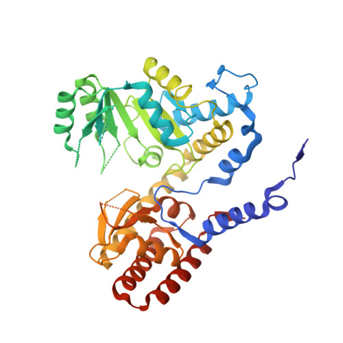
    Serine hydroxymethyltransferase (SHMT) is a pyridoxal-5'-phosphate (PLP)-dependent enzyme belonging to the fold type I superfamily, which catalyzes in vivo the reversible conversion of l-serine and tetrahydropteroylglutamate (H₄PteGlu) to glycine and 5,10-methylenetetrahydropteroylglutamate (5,10-CH₂-H₄PteGlu). The SHMT from the psychrophilic bacterium Psychromonas ingrahamii (piSHMT) had been recently purified and characterized. This enzyme was shown to display catalytic and stability properties typical of psychrophilic enzymes, namely high catalytic activity at low temperature and thermolability. To gain deeper insights into the structure-function relationship of piSHMT, the three-dimensional structure of its apo form was determined by X-ray crystallography. Homology modeling techniques were applied to build a model of the piSHMT holo form. Comparison of the two forms unraveled the conformation modifications that take place when the apo enzyme binds its cofactor. Our results show that the apo form is in an "open" conformation and possesses four (or five, in chain A) disordered loops whose electron density is not visible by X-ray crystallography. These loops contain residues that interact with the PLP cofactor and three of them are localized in the major domain that, along with the small domain, constitutes the single subunit of the SHMT homodimer. Cofactor binding triggers a rearrangement of the small domain that moves toward the large domain and screens the PLP binding site at the solvent side. Comparison to the mesophilic apo SHMT from Salmonella typhimurium suggests that the backbone conformational changes are wider in psychrophilic SHMT.


  • Organizational Affiliation
    • Dipartimento di Scienze Biochimiche "A. Rossi Fanelli", Università La Sapienza, 00185, Roma, Italy.

Macromolecule Content 

  • Total Structure Weight: 93.84 kDa 
  • Atom Count: 6,481 
  • Modeled Residue Count: 765 
  • Deposited Residue Count: 858 
  • Unique protein chains: 1

Macromolecules

Find similar proteins by:|  3D Structure
Entity ID: 1
MoleculeChains  Sequence LengthOrganismDetailsImage
Serine hydroxymethyltransferase
A, B
429Psychromonas ingrahamii 37Mutation(s): 0 
Gene Names: glyAPing_1438
EC: 2.1.2.1
UniProt
Find proteins for A1SUU0 (Psychromonas ingrahamii (strain DSM 17664 / CCUG 51855 / 37))
Explore A1SUU0 
Go to UniProtKB:  A1SUU0
Entity Groups
Sequence Clusters30% Identity50% Identity70% Identity90% Identity95% Identity100% Identity
UniProt GroupA1SUU0
Sequence Annotations
Expand
Reference Sequence

Small Molecules

Ligands 3 Unique
IDChains Name / Formula / InChI Key2D Diagram3D Interactions
SO4

Query on SO4


Download:Ideal Coordinates CCD File
C [auth A],
D [auth A],
E [auth A],
F [auth A],
N [auth B]
SULFATE ION
O4 S
QAOWNCQODCNURD-UHFFFAOYSA-L
GOL

Query on GOL


Download:Ideal Coordinates CCD File
J [auth A]
K [auth A]
L [auth A]
M [auth A]
O [auth B]
J [auth A],
K [auth A],
L [auth A],
M [auth A],
O [auth B],
P [auth B],
Q [auth B]
GLYCEROL
C3 H8 O3
PEDCQBHIVMGVHV-UHFFFAOYSA-N
CL

Query on CL


Download:Ideal Coordinates CCD File
G [auth A],
H [auth A],
I [auth A]
CHLORIDE ION
Cl
VEXZGXHMUGYJMC-UHFFFAOYSA-M

Experimental Data & Validation

Experimental Data

  • Method: X-RAY DIFFRACTION
  • Resolution: 1.85 Å
  • R-Value Free:  0.212 (Depositor), 0.216 (DCC) 
  • R-Value Work:  0.169 (Depositor), 0.173 (DCC) 
  • R-Value Observed: 0.170 (Depositor) 
Space Group: C 1 2 1
Unit Cell:
Length ( Å )Angle ( ˚ )
a = 146.82α = 90
b = 64.86β = 125.49
c = 116.94γ = 90
Software Package:
Software NamePurpose
PHENIXrefinement

Structure Validation

View Full Validation Report


Entry History 

Deposition Data

Revision History  (Full details and data files)

  • Version 1.0: 2014-07-30
    Type: Initial release
  • Version 1.1: 2014-08-20
    Changes: Database references
  • Version 1.2: 2014-10-08
    Changes: Database references
  • Version 1.3: 2023-12-20
    Changes: Data collection, Database references, Refinement description