4P3C

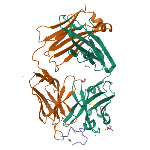
MT1-MMP:Fab complex (Form I)


Experimental Data Snapshot

  • Method: X-RAY DIFFRACTION
  • Resolution: 1.94 Å
  • R-Value Free: 0.200 
  • R-Value Work: 0.152 
  • R-Value Observed: 0.156 

Starting Model: experimental
View more details

wwPDB Validation   3D Report Full Report


This is version 1.6 of the entry. See complete history


Literature

Inhibition mechanism of membrane metalloprotease by an exosite-swiveling conformational antibody.

Udi, Y.Grossman, M.Solomonov, I.Dym, O.Rozenberg, H.Moreno, V.Cuniasse, P.Dive, V.Arroyo, A.G.Sagi, I.

(2015) Structure 23: 104-115

  • DOI: https://doi.org/10.1016/j.str.2014.10.012
  • Primary Citation of Related Structures:  
    4OUU, 4P3C, 4P3D, 4QXU

  • PubMed Abstract: 

    Membrane type 1 metalloprotease (MT1-MMP) is a membrane-anchored, zinc-dependent protease. MT1-MMP is an important mediator of cell migration and invasion, and overexpression of this enzyme has been correlated with the malignancy of various tumor types. Therefore, modulators of MT1-MMP activity are proposed to possess therapeutic potential in numerous invasive diseases. Here we report the inhibition mode of MT1-MMP by LEM-2/15 antibody, which targets a surface epitope of MT1-MMP. Specifically, the crystal structures of Fab LEM-2/15 in complex with the MT1-MMP surface antigen suggest that conformational swiveling of the enzyme surface loop is required for effective binding and consequent inhibition of MT1-MMP activity on the cell membrane. This inhibition mechanism appears to be effective in controlling active MT1-MMP in endothelial cells and at the leading edge of migratory cancer cells.


  • Organizational Affiliation

    Department of Biological Regulation, The Weizmann Institute of Science, Rehovot 76100, Israel.


Macromolecules
Find similar proteins by:  (by identity cutoff)  |  3D Structure
Entity ID: 1
MoleculeChains Sequence LengthOrganismDetailsImage
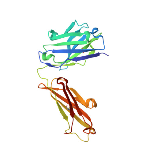
Heavy Chain Fab fragment of antibody LEM-2/15A [auth H]218Mus musculusMutation(s): 0 
Entity Groups  
Sequence Clusters30% Identity50% Identity70% Identity90% Identity95% Identity100% Identity
Sequence Annotations
Expand
  • Reference Sequence
Find similar proteins by:  (by identity cutoff)  |  3D Structure
Entity ID: 2
MoleculeChains Sequence LengthOrganismDetailsImage
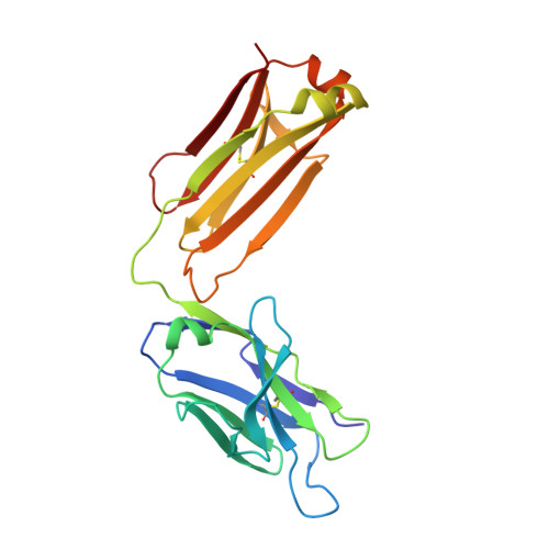
Light Chain Fab fragment of antibody LEM-2/15B [auth L]218Mus musculusMutation(s): 0 
Entity Groups  
Sequence Clusters30% Identity50% Identity70% Identity90% Identity95% Identity100% Identity
Sequence Annotations
Expand
  • Reference Sequence

Find similar proteins by:  Sequence   |   3D Structure  

Entity ID: 3
MoleculeChains Sequence LengthOrganismDetailsImage
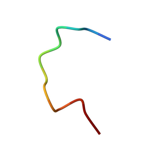
Matrix metalloproteinase-14C [auth M]13Homo sapiensMutation(s): 0 
Gene Names: MMP14
EC: 3.4.24.80
UniProt & NIH Common Fund Data Resources
Find proteins for P50281 (Homo sapiens)
Explore P50281 
Go to UniProtKB:  P50281
PHAROS:  P50281
GTEx:  ENSG00000157227 
Entity Groups  
Sequence Clusters30% Identity50% Identity70% Identity90% Identity95% Identity100% Identity
UniProt GroupP50281
Sequence Annotations
Expand
  • Reference Sequence
Small Molecules
Ligands 5 Unique
IDChains Name / Formula / InChI Key2D Diagram3D Interactions
EPE
Query on EPE

Download Ideal Coordinates CCD File 
E [auth H]4-(2-HYDROXYETHYL)-1-PIPERAZINE ETHANESULFONIC ACID
C8 H18 N2 O4 S
JKMHFZQWWAIEOD-UHFFFAOYSA-N
EDO
Query on EDO

Download Ideal Coordinates CCD File 
F [auth H]
G [auth H]
H
I [auth H]
J [auth H]
F [auth H],
G [auth H],
H,
I [auth H],
J [auth H],
N [auth L]
1,2-ETHANEDIOL
C2 H6 O2
LYCAIKOWRPUZTN-UHFFFAOYSA-N
ACT
Query on ACT

Download Ideal Coordinates CCD File 
K [auth H],
L [auth H],
O [auth L]
ACETATE ION
C2 H3 O2
QTBSBXVTEAMEQO-UHFFFAOYSA-M
CL
Query on CL

Download Ideal Coordinates CCD File 
D [auth H]CHLORIDE ION
Cl
VEXZGXHMUGYJMC-UHFFFAOYSA-M
MG
Query on MG

Download Ideal Coordinates CCD File 
M [auth L]MAGNESIUM ION
Mg
JLVVSXFLKOJNIY-UHFFFAOYSA-N
Experimental Data & Validation

Experimental Data

  • Method: X-RAY DIFFRACTION
  • Resolution: 1.94 Å
  • R-Value Free: 0.200 
  • R-Value Work: 0.152 
  • R-Value Observed: 0.156 
  • Space Group: P 2 21 21
Unit Cell:
Length ( Å )Angle ( ˚ )
a = 52.314α = 90
b = 80.975β = 90
c = 95.568γ = 90
Software Package:
Software NamePurpose
DNAdata collection
HKL-2000data scaling
PHASERphasing
PHENIXrefinement

Structure Validation

View Full Validation Report


Entry History 

Deposition Data

Revision History  (Full details and data files)

  • Version 1.0: 2014-12-17
    Type: Initial release
  • Version 1.1: 2015-01-14
    Changes: Database references
  • Version 1.2: 2015-01-21
    Changes: Database references
  • Version 1.3: 2015-02-18
    Changes: Refinement description
  • Version 1.4: 2017-09-27
    Changes: Advisory, Database references, Derived calculations, Other, Refinement description, Source and taxonomy
  • Version 1.5: 2023-09-27
    Changes: Data collection, Database references, Derived calculations, Refinement description
  • Version 1.6: 2024-10-30
    Changes: Structure summary