4P2Y | pdb_00004p2y

Crystal structure of the human RAGE ectodomain (fragment VC1C2) in complex with mouse S100A6


Experimental Data Snapshot

  • Method: X-RAY DIFFRACTION
  • Resolution: 2.30 Å
  • R-Value Free: 
    0.207 (Depositor), 0.216 (DCC) 
  • R-Value Work: 
    0.187 (Depositor), 0.194 (DCC) 
  • R-Value Observed: 
    0.188 (Depositor) 

Starting Models: experimental
View more details

wwPDB Validation 3D Report Full Report

Validation slider image for 4P2Y

This is version 1.4 of the entry. See complete history

Literature

The Structure of the RAGE:S100A6 Complex Reveals a Unique Mode of Homodimerization for S100 Proteins.

Yatime, L.Betzer, C.Jensen, R.K.Mortensen, S.Jensen, P.H.Andersen, G.R.

(2016) Structure 

  • DOI: https://doi.org/10.1016/j.str.2016.09.011
  • Primary Citation Related Structures: 
    4P2Y, 4YBH

  • PubMed Abstract: 

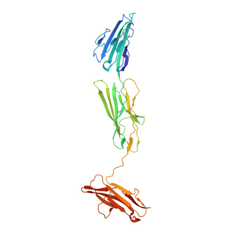
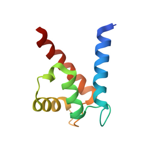
    S100 proteins are calcium-dependent regulators of homeostatic processes. Upon cellular response to stress, and notably during tumorigenesis, they relocalize to the extracellular environment where they induce pro-inflammatory signals by activating the receptor for advanced glycation end products (RAGE), thereby facilitating tumor growth and metastasis. Despite its importance in sustaining inflammation, the structural basis for RAGE-S100 crosstalk is still unknown. Here we report two crystal structures of the RAGE:S100A6 complex encompassing a full-length RAGE ectodomain. The structures, in combination with a comprehensive interaction analysis, suggest that the primary S100A6 binding site is formed by the RAGE C1 domain. Complex formation with S100A6 induces a unique dimeric conformation of RAGE that appears suited for signal transduction and intracellular effector recruitment. Intriguingly, S100A6 adopts a dimeric conformation radically different from all known S100 dimers. We discuss the physiological relevance of this non-canonical homodimeric form in vivo.


  • Organizational Affiliation
    • Department of Molecular Biology and Genetics, Aarhus University, Gustav Wieds Vej 10C, 8000 Aarhus, Denmark. Electronic address: laure.yatime@inserm.fr.

Macromolecule Content 

  • Total Structure Weight: 43.97 kDa 
  • Atom Count: 3,213 
  • Modeled Residue Count: 389 
  • Deposited Residue Count: 395 
  • Unique protein chains: 2

Macromolecules

Find similar proteins by:|  3D Structure
Entity ID: 1
MoleculeChains  Sequence LengthOrganismDetailsImage
Advanced glycosylation end product-specific receptor304Homo sapiensMutation(s): 0 
Gene Names: AGERRAGE
UniProt & NIH Common Fund Data Resources
Find proteins for Q15109 (Homo sapiens)
Explore Q15109 
Go to UniProtKB:  Q15109
PHAROS:  Q15109
GTEx:  ENSG00000204305 
Entity Groups
Sequence Clusters30% Identity50% Identity70% Identity90% Identity95% Identity100% Identity
UniProt GroupQ15109
Sequence Annotations
Expand
Reference Sequence
Find similar proteins by:|  3D Structure
Entity ID: 2
MoleculeChains  Sequence LengthOrganismDetailsImage
Protein S100-A691Mus musculusMutation(s): 0 
Gene Names: S100a6Cacy
UniProt & NIH Common Fund Data Resources
Find proteins for P14069 (Mus musculus)
Explore P14069 
Go to UniProtKB:  P14069
IMPC:  MGI:1339467
Entity Groups
Sequence Clusters30% Identity50% Identity70% Identity90% Identity95% Identity100% Identity
UniProt GroupP14069
Sequence Annotations
Expand
Reference Sequence

Small Molecules

Ligands 4 Unique
IDChains Name / Formula / InChI Key2D Diagram3D Interactions
ZN

Query on ZN


Download:Ideal Coordinates CCD File
C [auth A]
D [auth A]
E [auth A]
F [auth A]
R [auth B]
C [auth A],
D [auth A],
E [auth A],
F [auth A],
R [auth B],
V [auth B]
ZINC ION
Zn
PTFCDOFLOPIGGS-UHFFFAOYSA-N
ACT

Query on ACT


Download:Ideal Coordinates CCD File
L [auth A]
M [auth A]
N [auth A]
O [auth A]
P [auth A]
L [auth A],
M [auth A],
N [auth A],
O [auth A],
P [auth A],
Q [auth A],
W [auth B]
ACETATE ION
C2 H3 O2
QTBSBXVTEAMEQO-UHFFFAOYSA-M
CA

Query on CA


Download:Ideal Coordinates CCD File
T [auth B],
U [auth B]
CALCIUM ION
Ca
BHPQYMZQTOCNFJ-UHFFFAOYSA-N
CL

Query on CL


Download:Ideal Coordinates CCD File
G [auth A]
H [auth A]
I [auth A]
J [auth A]
K [auth A]
G [auth A],
H [auth A],
I [auth A],
J [auth A],
K [auth A],
S [auth B]
CHLORIDE ION
Cl
VEXZGXHMUGYJMC-UHFFFAOYSA-M

Experimental Data & Validation

Experimental Data

  • Method: X-RAY DIFFRACTION
  • Resolution: 2.30 Å
  • R-Value Free:  0.207 (Depositor), 0.216 (DCC) 
  • R-Value Work:  0.187 (Depositor), 0.194 (DCC) 
  • R-Value Observed: 0.188 (Depositor) 
Space Group: I 2 2 2
Unit Cell:
Length ( Å )Angle ( ˚ )
a = 76.78α = 90
b = 113.39β = 90
c = 140.32γ = 90
Software Package:
Software NamePurpose
XDSdata scaling
XSCALEdata scaling
PDB_EXTRACTdata extraction
PHASERphasing
PHENIXrefinement

Structure Validation

View Full Validation Report


Entry History 

Deposition Data

Revision History  (Full details and data files)

  • Version 1.0: 2015-03-11
    Type: Initial release
  • Version 1.1: 2016-11-23
    Changes: Database references
  • Version 1.2: 2017-11-22
    Changes: Database references, Refinement description
  • Version 1.3: 2023-09-27
    Changes: Data collection, Database references, Derived calculations, Refinement description
  • Version 1.4: 2024-10-23
    Changes: Structure summary