4P2F | pdb_00004p2f

Monomeric form of a single mutant (F363R) of Mycobacterial Adenylyl cyclase Rv1625c


Experimental Data Snapshot

  • Method: X-RAY DIFFRACTION
  • Resolution: 2.05 Å
  • R-Value Free: 
    0.233 (Depositor), 0.234 (DCC) 
  • R-Value Work: 
    0.179 (Depositor), 0.179 (DCC) 
  • R-Value Observed: 
    0.182 (Depositor) 

Starting Model: experimental
View more details

wwPDB Validation 3D Report Full Report

Validation slider image for 4P2F

This is version 1.3 of the entry. See complete history

Literature

New structural forms of a mycobacterial adenylyl cyclase Rv1625c.

Barathy, D.Mattoo, R.Visweswariah, S.Suguna, K.

(2014) IUCrJ 1: 338-348

  • DOI: https://doi.org/10.1107/S2052252514016741
  • Primary Citation Related Structures: 
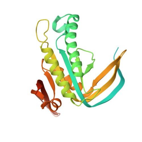
    4P2F, 4P2M, 4P2X

  • PubMed Abstract: 

    Rv1625c is one of 16 adenylyl cyclases encoded in the genome of Mycobacterium tuberculosis. In solution Rv1625c exists predominantly as a monomer, with a small amount of dimer. It has been shown previously that the monomer is active and the dimeric fraction is inactive. Both fractions of wild-type Rv1625c crystallized as head-to-head inactive domain-swapped dimers as opposed to the head-to-tail dimer seen in other functional adenylyl cyclases. About half of the molecule is involved in extensive domain swapping. The strain created by a serine residue located on a hinge loop and the crystallization condition might have led to this unusual domain swapping. The inactivity of the dimeric form of Rv1625c could be explained by the absence of the required catalytic site in the swapped dimer. A single mutant of the enzyme was also generated by changing a phenylalanine predicted to occur at the functional dimer interface to an arginine. This single mutant exists as a dimer in solution but crystallized as a monomer. Analysis of the structure showed that a salt bridge formed between a glutamate residue in the N-terminal segment and the mutated arginine residue hinders dimer formation by pulling the N-terminal region towards the dimer interface. Both structures reported here show a change in the dimerization-arm region which is involved in formation of the functional dimer. It is concluded that the dimerization arm along with other structural elements such as the N-terminal region and certain loops are vital for determining the oligomeric nature of the enzyme, which in turn dictates its activity.


  • Organizational Affiliation
    • Molecular Biophysics Unit, Indian Institute of Science , C. V. Raman Avenue, Bangalore, Karnataka 560 012, India.

Macromolecule Content 

  • Total Structure Weight: 31.31 kDa 
  • Atom Count: 1,631 
  • Modeled Residue Count: 189 
  • Deposited Residue Count: 273 
  • Unique protein chains: 1

Macromolecules

Find similar proteins by:|  3D Structure
Entity ID: 1
MoleculeChains  Sequence LengthOrganismDetailsImage
Adenylate cyclase273Mycobacterium tuberculosisMutation(s): 0 
Gene Names: cyaRv1625cMT1661MTCY01B2.17c
EC: 4.6.1.1
UniProt
Find proteins for P9WQ35 (Mycobacterium tuberculosis (strain ATCC 25618 / H37Rv))
Explore P9WQ35 
Go to UniProtKB:  P9WQ35
Entity Groups
Sequence Clusters30% Identity50% Identity70% Identity90% Identity95% Identity100% Identity
UniProt GroupP9WQ35
Sequence Annotations
Expand
Reference Sequence

Small Molecules

Ligands 3 Unique
IDChains Name / Formula / InChI Key2D Diagram3D Interactions
PEG

Query on PEG


Download:Ideal Coordinates CCD File
T [auth A]DI(HYDROXYETHYL)ETHER
C4 H10 O3
MTHSVFCYNBDYFN-UHFFFAOYSA-N
GOL

Query on GOL


Download:Ideal Coordinates CCD File
U [auth A]GLYCEROL
C3 H8 O3
PEDCQBHIVMGVHV-UHFFFAOYSA-N
EDO

Query on EDO


Download:Ideal Coordinates CCD File
B [auth A]
C [auth A]
D [auth A]
E [auth A]
F [auth A]
B [auth A],
C [auth A],
D [auth A],
E [auth A],
F [auth A],
G [auth A],
H [auth A],
I [auth A],
J [auth A],
K [auth A],
L [auth A],
M [auth A],
N [auth A],
O [auth A],
P [auth A],
Q [auth A],
R [auth A],
S [auth A]
1,2-ETHANEDIOL
C2 H6 O2
LYCAIKOWRPUZTN-UHFFFAOYSA-N

Experimental Data & Validation

Experimental Data

  • Method: X-RAY DIFFRACTION
  • Resolution: 2.05 Å
  • R-Value Free:  0.233 (Depositor), 0.234 (DCC) 
  • R-Value Work:  0.179 (Depositor), 0.179 (DCC) 
  • R-Value Observed: 0.182 (Depositor) 
Space Group: P 21 21 21
Unit Cell:
Length ( Å )Angle ( ˚ )
a = 29.89α = 90
b = 67.37β = 90
c = 101.55γ = 90
Software Package:
Software NamePurpose
REFMACrefinement

Structure Validation

View Full Validation Report


Entry History 

Deposition Data

Revision History  (Full details and data files)

  • Version 1.0: 2014-09-17
    Type: Initial release
  • Version 1.1: 2014-10-22
    Changes: Database references
  • Version 1.2: 2017-11-01
    Changes: Author supporting evidence, Derived calculations, Other, Source and taxonomy, Structure summary
  • Version 1.3: 2023-09-27
    Changes: Data collection, Database references, Refinement description