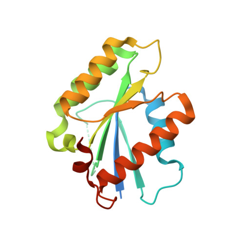
4P0T | pdb_00004p0t

Crystal structure of human centromere protein M


Experimental Data Snapshot

  • Method: X-RAY DIFFRACTION
  • Resolution: 1.49 Å
  • R-Value Free: 
    0.164 (Depositor), 0.161 (DCC) 
  • R-Value Work: 
    0.124 (Depositor), 0.125 (DCC) 
  • R-Value Observed: 
    0.127 (Depositor) 

wwPDB Validation 3D Report Full Report

Validation slider image for 4P0T

This is version 1.2 of the entry. See complete history. 

Literature

The pseudo GTPase CENP-M drives human kinetochore assembly.

Basilico, F., Maffini, S., Weir, J.R., Prumbaum, D., Rojas, A.M., Zimniak, T., De Antoni, A., Jeganathan, S., Voss, B., van Gerwen, S., Krenn, V., Massimiliano, L., Valencia, A., Vetter, I.R., Herzog, F., Raunser, S., Pasqualato, S., Musacchio, A.

(2014) Elife 3: e02978-e02978

  • DOI: https://doi.org/10.7554/eLife.02978
  • Primary Citation Related Structures: 
    4P0T

  • PubMed Abstract: 

    Kinetochores, multi-subunit complexes that assemble at the interface with centromeres, bind spindle microtubules to ensure faithful delivery of chromosomes during cell division. The configuration and function of the kinetochore-centromere interface is poorly understood. We report that a protein at this interface, CENP-M, is structurally and evolutionarily related to small GTPases but is incapable of GTP-binding and conformational switching. We show that CENP-M is crucially required for the assembly and stability of a tetramer also comprising CENP-I, CENP-H, and CENP-K, the HIKM complex, which we extensively characterize through a combination of structural, biochemical, and cell biological approaches. A point mutant affecting the CENP-M/CENP-I interaction hampers kinetochore assembly and chromosome alignment and prevents kinetochore recruitment of the CENP-T/W complex, questioning a role of CENP-T/W as founder of an independent axis of kinetochore assembly. Our studies identify a single pathway having CENP-C as founder, and CENP-H/I/K/M and CENP-T/W as CENP-C-dependent followers.DOI: http://dx.doi.org/10.7554/eLife.02978.001.


  • Organizational Affiliation: 
    • Department of Mechanistic Cell Biology, Max Planck Institute of Molecular Physiology, Dortmund, Germany Department of Experimental Oncology, European Institute of Oncology, Milan, Italy.

Macromolecule Content 

  • Total Structure Weight: 38.58 kDa 
  • Atom Count: 2,610 
  • Modeled Residue Count: 298 
  • Deposited Residue Count: 352 
  • Unique protein chains: 1

Macromolecules

Find similar proteins by:|  3D Structure
Entity ID: 1
MoleculeChains  Sequence LengthOrganismDetailsImage
Centromere protein M
A, B
176Homo sapiensMutation(s): 0 
Gene Names: CENPM, C22orf18, ICEN39, PANE1
UniProt & NIH Common Fund Data Resources
Find proteins for Q9NSP4 (Homo sapiens)
Explore Q9NSP4 
Go to UniProtKB:  Q9NSP4
PHAROS:  Q9NSP4
GTEx:  ENSG00000100162 
Entity Groups
Sequence Clusters30% Identity50% Identity70% Identity90% Identity95% Identity100% Identity
UniProt GroupQ9NSP4
Sequence Annotations
Expand
Reference Sequence

Small Molecules

Ligands 1 Unique
IDChains Name / Formula / InChI Key2D Diagram3D Interactions
GOL

Query on GOL


Download:Ideal Coordinates CCD File
C [auth B]GLYCEROL
C3 H8 O3
PEDCQBHIVMGVHV-UHFFFAOYSA-N

Experimental Data & Validation

Experimental Data

  • Method: X-RAY DIFFRACTION
  • Resolution: 1.49 Å
  • R-Value Free:  0.164 (Depositor), 0.161 (DCC) 
  • R-Value Work:  0.124 (Depositor), 0.125 (DCC) 
  • R-Value Observed: 0.127 (Depositor) 
Space Group: P 3
Unit Cell:
Length ( Å )Angle ( ˚ )
a = 104.03α = 90
b = 104.03β = 90
c = 33.56γ = 120
Software Package:
Software NamePurpose
PHENIXrefinement
PHENIXphasing
Cootmodel building
xia2data reduction

Structure Validation

View Full Validation Report


Entry History 

Deposition Data

Revision History  (Full details and data files)

  • Version 1.0: 2014-07-09
    Type: Initial release
  • Version 1.1: 2014-07-23
    Changes: Database references
  • Version 1.2: 2024-05-08
    Changes: Data collection, Database references