4P09 | pdb_00004p09

Crystal structure of HOIP PUB domain


Experimental Data Snapshot

  • Method: X-RAY DIFFRACTION
  • Resolution: 1.70 Å
  • R-Value Free: 
    0.230 (Depositor), 0.250 (DCC) 
  • R-Value Work: 
    0.181 (Depositor) 
  • R-Value Observed: 
    0.184 (Depositor) 

wwPDB Validation 3D Report Full Report

Validation slider image for 4P09

This is version 1.4 of the entry. See complete history

Literature

Binding of OTULIN to the PUB domain of HOIP controls NF-kappa B signaling.

Schaeffer, V.Akutsu, M.Olma, M.H.Gomes, L.C.Kawasaki, M.Dikic, I.

(2014) Mol Cell 54: 349-361

  • DOI: https://doi.org/10.1016/j.molcel.2014.03.016
  • Primary Citation Related Structures: 
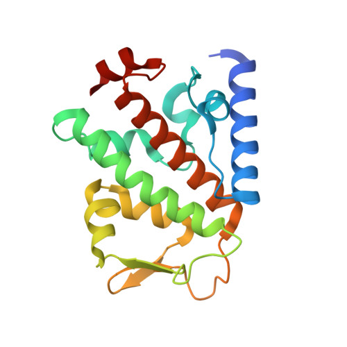
    4P09, 4P0A, 4P0B

  • PubMed Abstract: 

    Linear ubiquitin chains are implicated in the regulation of the NF-κB pathway, immunity, and inflammation. They are synthesized by the LUBAC complex containing the catalytic subunit HOIL-1-interacting protein (HOIP) and are disassembled by the linear ubiquitin-specific deubiquitinase OTULIN. Little is known about the regulation of these opposing activities. Here we demonstrate that HOIP and OTULIN interact and act as a bimolecular editing pair for linear ubiquitin signals in vivo. The HOIP PUB domain binds to the PUB interacting motif (PIM) of OTULIN and the chaperone VCP/p97. Structural studies revealed the basis of high-affinity interaction with the OTULIN PIM. The conserved Tyr56 of OTULIN makes critical contacts with the HOIP PUB domain, and its phosphorylation negatively regulates this interaction. Functionally, HOIP binding to OTULIN is required for the recruitment of OTULIN to the TNF receptor complex and to counteract HOIP-dependent activation of the NF-κB pathway.


  • Organizational Affiliation
    • Institute of Biochemistry II, Goethe University Faculty of Medicine, 60590 Frankfurt am Main, Germany.

Macromolecule Content 

  • Total Structure Weight: 20.77 kDa 
  • Atom Count: 1,599 
  • Modeled Residue Count: 177 
  • Deposited Residue Count: 184 
  • Unique protein chains: 1

Macromolecules

Find similar proteins by:|  3D Structure
Entity ID: 1
MoleculeChains  Sequence LengthOrganismDetailsImage
E3 ubiquitin-protein ligase RNF31184Homo sapiensMutation(s): 0 
Gene Names: RNF31ZIBRA
EC: 6.3.2 (PDB Primary Data), 2.3.2.31 (UniProt)
UniProt & NIH Common Fund Data Resources
Find proteins for Q96EP0 (Homo sapiens)
Explore Q96EP0 
Go to UniProtKB:  Q96EP0
PHAROS:  Q96EP0
GTEx:  ENSG00000092098 
Entity Groups
Sequence Clusters30% Identity50% Identity70% Identity90% Identity95% Identity100% Identity
UniProt GroupQ96EP0
Sequence Annotations
Expand
Reference Sequence

Experimental Data & Validation

Experimental Data

  • Method: X-RAY DIFFRACTION
  • Resolution: 1.70 Å
  • R-Value Free:  0.230 (Depositor), 0.250 (DCC) 
  • R-Value Work:  0.181 (Depositor) 
  • R-Value Observed: 0.184 (Depositor) 
Space Group: P 41 21 2
Unit Cell:
Length ( Å )Angle ( ˚ )
a = 47.92α = 90
b = 47.92β = 90
c = 152.71γ = 90
Software Package:
Software NamePurpose
PHENIXrefinement
PDB_EXTRACTdata extraction

Structure Validation

View Full Validation Report


Entry History 

Deposition Data

Revision History  (Full details and data files)

  • Version 1.0: 2014-05-07
    Type: Initial release
  • Version 1.1: 2014-05-21
    Changes: Data collection
  • Version 1.2: 2014-10-01
    Changes: Database references
  • Version 1.3: 2017-11-22
    Changes: Data collection, Database references, Derived calculations, Other, Refinement description, Source and taxonomy, Structure summary
  • Version 1.4: 2023-12-27
    Changes: Data collection, Database references, Refinement description